Download
1 / 22
220 likes | 258 Views
Surface Science at the Soft X-ray Beamline. Anton Tadich Soft X-ray Spectroscopy eamline. Soft X-ray Spectroscopy. Soft X-ray Region. X-ray Interaction with matter. F or Soft X -ray Energies : X-ray absorption ( “electron absorbs photon”) probability dominates by orders of magnitude.

E N D
Surface Scienceat the Soft X-ray Beamline Anton Tadich Soft X-ray Spectroscopy eamline
Soft X-ray Spectroscopy Soft X-ray Region • X-ray Interaction with matter • For Soft X-ray Energies: • X-ray absorption (“electron absorbs photon”) probability dominates by orders of magnitude • We offer two main techniques: • Near Edge X-ray Absorption Fine Structure (NEXAFS) • Soft X-ray Photoelectron Spectroscopy (SXPS) Courtesy: J.H Hubbell et al. J. Phys. Chem .Ref. Data 9, (1023), 1980
NEXAFS Spectroscopy X-ray absorption X-ray Absorption Spectroscopy • Measure the x-ray absorption of the sample as the x-ray energy is tuned across the “edge energy” of the core level hn K Edge Near Edge X-ray Absorption Spectroscopy (NEXAFS) • Extended X-ray Absorption Fine Structure (EXAFS) • Local probe of structure around emitter using photoelectron wave • Interference between outgoing electron wave and backscattered wave off neighboring atoms • Probe transitions to unoccupied, bound states • Sensitive to local chemical environment, bond geometry http://upload.wikimedia.org/wikipedia/commons/thumb/c/c2/NEXAFS_EXAFS_schematic.svg/613px-NEXAFS_EXAFS_schematic.svg.png
Molecular orientation using NEXAFS Molecular Orientation with NEXAFS • NEXAFS with polarised light is a powerful tool for determining the orientation of molecular orbitals • Polarised soft x-rays act as a “search” light for unoccupied orbitals aligned with the E vector J. Stohr SSRL
Example: Melamine on graphene Optimised DFT adsorption geometry • Context: Using graphene as small molecule sensor • C K- edge and N K- edge NEXAFS data suggest a flat adsorption geometry up to 3.6ML • Amino p* angle dependence indicates 8°tilt angle from plane Courtesy J Cervenka, University of Melbourne
Soft X-ray Photoelectron Spectroscopy Electron Binding Energy • Electrons in atomic core shells (1s, 2s, 2p,etc) are bound to the nucleus with element specific binding energies http://xdb.lbl.gov/Section1/Table_1-1a.htm http://www.ifw-dresden.de/institutes/ikm/organisation/dep-31/methods/x-ray-photoelectron-spectroscopy-xps/xps2.jpg
Soft X-ray Photoelectron Spectroscopy • The Photoemission Process • With sufficient photon energy, electrons from occupied core levels can be liberated and detected with an electron spectrometer • The kinetic energy of the electron yields its corresponding binding energy EB via the equation: • Ekin= hn – EB – f • (where f represents the work function of spectrometer) hn Ekin f EB
Creating a 2D hole gas on diamond with C60F48 • Soft X-ray Photoemission: X-ray in – Electron out technique • Probes chemical and charge environment of molecules on the surface C1s @ 330eV SXR light e- e- e- e- • Ekin= hn – EB – f Ionised (doping) and neutral (non-doping) C60F48 components are resolved!
XPS WITH SR: ADVANTAGES 1. Cross Section Optimization C1s Excitation Cross Section • The photoionisation cross section for a given shell (e.g 1s or K) exhibits a rapid increase, followed by a smooth decrease, at the threshold energy SR @ 600eV • For lab based X-ray source energies (e.g Al-Ka 1486.6eV), the cross-section is quite low for light, low Z elements Cross Section (Mbarn) Lab XPS @1486.6eV One can lower the x-ray energy to obtain an order or more magnitude in excitation probability Photon Energy (eV) http://ulisse.elettra.trieste.it/services/elements/WebElements.html 10
SXPS: surface sensitivity Electron Mean Free Path • XPS derives its surface sensitivity from the fact that photoelectrons and Auger electrons possess extremely short mean free paths (l) hn I = I0e-d/l d IO I0 http://www.philiphofmann.net/surflec/fig3_2.gif • 95% of photoelectrons have scattered within 3l from the surface
XPS WITH SR: ADVANTAGES 2. Tuning The Core Level Kinetic Energy • Inelastic scattering => most of the signal comes from a few MFP of the surface. • XPS is extremely surface sensitive, http://www.philiphofmann.net/surflec/fig3_2.gif With SR: one can “tune” the KE of a photoelectron to obtain depth information Qualitative (Easy) Quantitative (Harder)
Black Phosphorus Oxidation Cleave in Vacuum -> measure Expose to air -> measure
Oxide Peaks O1s=531.62 eV O1s=533.24 eV O1s=531.7 eV O1s=533.49 eV How we are beginning to interpret the data Literature values for Phosphite 531.8 eV and 533.3 eV J. Non-cryst. Solids 160, 73 (1993) 531.5 eV and 533.3 eV Phys. Chem. Glass. 36, 247 (1996) The lower BE O1s corresponds to bridging oxygen and higher BE O1s to non-bridging oxygen in NaPO3. .
Black Phosphorus Oxide Peaks P2p3/2=130.06 eV P2p1/2=130.94 eV P2p3/2=130.17 eV P2p1/2=131.04 eV Oxide Thickness P2p3/2=130.58 eV P2p1/2=131.52 eV POxide1=132.65 POxide2=134.42 (PO3) 0.24nm P2p3/2=130.1 eV P2p1/2=130.95 eV P2p3/2=130.16 eV P2p1/2=131.01 eV P2p3/2=130.64 eV P2p1/2=131.58 eV POxide1=132.8 POxide2=134.65 (PO3) 0.43nm
hn = 180 eV 350eV 800eV This peak related to surface species
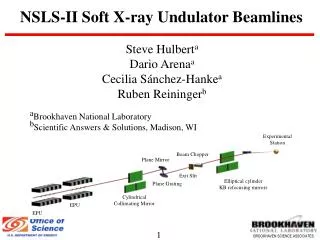
The Soft X-ray Endstation Multi purpose Ultra High Vacuum (UHV) endstation dedicated for XPS and NEXAFS • Key Features • Multiple NEXAFS detectors (PEY, TFY) • High resolution electron spectrometer • Multifunction preparation chamber • User friendly sample transfer • Crystal cleaving chamber • Inert atmosphere “glovebox” • Electron flood gun for insulators SAMPLE ENDSTATION IMAGE
Main Detectors Photoemission NEXAFS • SPECS Phoibos 150 hemispherical analyser • 150mm mean radius • 9 channeltron detector • K.E up to 3.5keV. DE = 141meV @ 10eV pass • Various Lens modes • Total electron yield (sample current) • Retarding Grid Analyzer(Partial Electron Yield or Total Fluorescence Yield) • Channeltron (Partial Electron yield) • Simultaneous bulk (<100nm) and surface (<10nm) NEXAFS
PREPARATION/CHARACTERISATION CHAMBER A wide range of sample preparation and characterisation options: • Residual Gas Analyser (to 300amu) • Electron beam evaporator (to >3000C) • Wide range effusion evaporator (200C to 1400C) • Organic Material Evaporator (RT to 300C) • Medium Temp Evaporator (300C to 800C) • Low Energy Electron Diffraction (LEED) • Quartz crystal microbalance • Argon ion sputtering 0.1 – 5keV • Heating/cooling of sample: -160C to 1200C • Gas Dosing (up to 10-6 mbar) • Cleaving of layered materials • 4-point conductivity probe (basic elec. meas.) • Kelvin Probe (Alt. work function measurement)
Sample Requirements Samples Size Requirements • Samples must fit on 25mm diameter disc • Sample height must be no more than 3 -4mm Form of Samples • Wafers, powders, crystals, liquids (ionic), minerals, polymers. • Samples must be UHV Compatible!!! Need to maintain < 10-9 mbar during measurement Also…. • Multiple samples possible per holder • Introduction time of single holder to system ~ 2 hours
Information for new users Applying as a New User The Beamtime • Merit based selection for beamtime • Contact beamline scientists for advice on experiment proposal! • Dedicated Beamline Scientist as “Local Contact”! • 4 to 6 days beamtime, depending on experiment and user skill • 1 day spent training without beam on the endstation • At least 1-2 days with beam before “real” data starts being taken Pre beamtime • A basic knowledge of photoelectron/Auger electron spectroscopy, and NEXAFS, goes a long way toward a successful experiment • Contact beamline staff regarding: samples, experimental plan, people,…
Information for new users NEXAFS Proposals XPS Proposals • Will need to demonstrate that you seek more than just “what’s there” • Will need to justify why a lab based XPS system is not suitable (surface sensitivity, cross section, resonance arguments) • Insulators: tend to be quite difficult, lineshape not good even with flood gun, fitting problematic • NEXAFS is more difficult to interpret than XPS, less literature on systems • Reference materials (e.g coordination chemistry, functional groups) vital • Insulators: very doable, more so than XPS • Carbon NEXAFS: Add extra day of learning, especially for dilute systems. Email: softxray@synchrotron.org.au Thank You!