Download
1 / 81
810 likes | 876 Views
Explore the intricate details of eye anatomy and physiology, from external structures to visual pathways. Learn about diagnostic tests, common disorders like cataract and glaucoma, and essential nursing care for eye surgeries.

E N D
NEUROSENSORY SYSTEM
ANATOMY & PHYSIOLOGY OF EYES EXTERNAL STRUCTURES • EYELIDS • CONJUNCTIVA • PALBEBRAL • BULBAR • LACRIMAL APPARATUS • LACRIMAL GLAND, DUCTS & PASSAGES • 6 EXTRAOCULAR MUSCLES • Levatorpalpebrae muscle
ANATOMY & PHYSIOLOGYEYES • ORBIT • EYEBALL : 3 LAYERS: • OUTER • SCLERA • CORNEA • MIDDLE • CHOROID • CILIARY BODY • IRIS • INNER • RODS • SENSITIVE TO LIGHT • PERIPHERAL VISION • CONES • FINE • DESCRIMINATION • COLOR VSION
ANATOMY & PHYSIOLOGYEYES • LENS – FOCUS IMAGE • FLUIDS OF THE EYE: • AQUEOUS HUMOR • ANTERIOR & POSTERIOR CHAMBERS • ANTERIOR EYE CAVITY • NUTRIENTS TO LENS & CORNEA • INTRAOCULAR PRESSURE MAINTENANCE • 20-25 mmHg • VITREOUS HUMOR • POSTERIOR EYE CAVITY • TRANSPARENCY & FORM OF THE EYE
VISUAL PATHWAYS RETINA OPTIC NERVE OPTIC CHIASM OPTIC TRACT OCCIPITAL LOBE
Physical Examination-EYE • VISUAL ACUITY : SNELLEN’S CHART • VISUAL FIELDS: PERIMETRY • EXTERNAL STRUCTURES • POSITION & ALIGNMENT OF EYES • PUPILS (PERRLA) • EXTRAOCULAR MOVEMENTS • PARALYSIS • NYSTAGMUS • CORNEAL REFLEX
DIAGNOSTIC TESTS • SNELLEN • OPHTHALMOSCOPE • BIOMICROSCOPE / SLITLAMP • EXAMINE THE ANTERIOR SEGMENT OF THE EYE • TONOMETER • 14-20 mmHg • BJERRUM’S TANGENT SCREEN • CENTRAL FIELD OF VISION • ISHIHARA COLOR PLATE TEST • IDENTIFY 3 PRIMARY COLORS • GONIOSCOPY • ANGLE OF ANTERIOR CHAMBER
PLANNING FOR HEALTH PROMOTION • PERSISTENT REDNESS • CONTINUED DISCOMFORT & PAIN ESP • FOLLOWING INJURY • CHILDREN: CROSSING OF EYES • BLURRED VISION/ SPOTS BEFORE THE EYES • GROWTH ON THE EYE/ OPACITIES • CONTINUAL DISCHARGE, CRUSTING OR • TEARING • PUPIL IRREGULARITIES CARE OF THE EYES • EYEDROPS, DISCOURAGED • PRINTED MATTER: 14 INCHES AWAY • TV: 10-12 FT AWAY • READ WITH ILLUMINATION: 100-150 WATTS • LIGHT FROM BEHIND • TEACH ABOUT DANGER SIGNALS OF VISUAL DISORDER
DISORDERS - EYE • INJURIES & TRAUMA • INFECTIONS • CATARACT • GLAUCOMA • DETACHMENT OF THE RETINA • REFRACTIVE ERRORS
INJURIES & TRAUMA EMERGENCY: • TREAT THE PATIENT, LEAVE THE EYE ALONE, EXCEPT IN CHEMICAL INJURY - FLUSH EYES STAT • FOREIGN BODIES: FLUSH WITH WATER FOR 15 MIN WHILE GOING TO THE DOCTOR; DON’T TOUCH CORNEA
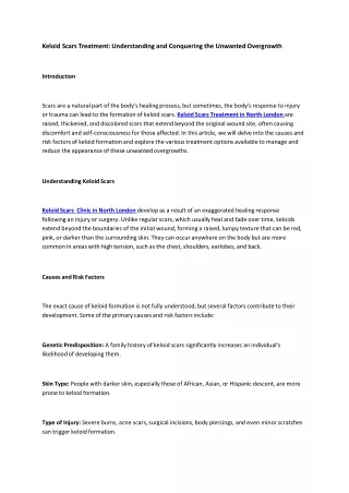
INFECTIONS • HORDEOLUM/ STY -Zeis gland in the follicle • CHALAZION –meibomian glands • CONJUNCTIVITIS – pink eye • bacterial infection, allergy, trauma • UVEITIS - iris • KERATITIS - cornea • PTERYGIUM – triangular fold • From white of the eye to the cornea
CATARACT • Opacity of the lens & its capsule which interferes with transparency S/SX: • Dimness in visual acuity • Rapid & marked refraction error CLASSIFICATION: • Primary/ senile • Secondary/ traumatic • Congenital
Treatment • Replacement of the intra ocular lens • Commonly done by phakoemulsification technique
EYE SURGERY NURSING CARE PRE-OP • Orient to new environment • Teach deep breathing & how to close eyes without squeezing • Eye antibiotics preop • Mydiatrics if ordered
EYE SURGERY NURSING CARE POST-OP • Reorient patient to his surroundings • Prevent increase in IOP & stress on the suture line
Contd…. • ACTIVITIES THAT INCREASE IOP: • Coughing • Vomiting • Bending • Stooping • Promote comfort of the patient: mild analgesic to control pain
EYE SURGERY • COMPLICATIONS: • NAUSEA & VOMITING • Antiemetics • Cold compress • HEMORRHAGE • Sudden pain of the eye • PROLAPSE OF THE IRIS • Most common postop complication • Can precipitate glaucoma NURSING CARE POST-OP • Observe & treat complications • Promote the rehab of the patient • Encourage the patient to become • independent- walk with him when he first • become ambulatory • Health teachings
EYE SURGERY HEALTH TEACHINGS: • 1-4 wks : dark glasses; temporary corrective lenses • 6-8 wks: permanent lenses • It will take time to learn distances & climb stairs • Color slightly changed • Use one eye at a time unless with contact lens • Decreased peripheral vision
GLAUCOMA • INCREASED IOP • PROGRESSIVE LOSS OF PERIPHERAL VISION CAUSE: OBSTRUCTION TO CIRCULATION OF AQUEOUS HUMOR TYPES: • CHRONIC/ SIMPLE/ OPEN-ANGLE • ACUTE ANGLE CLOSURE • Congenital • Secondary – trauma, uveitis, postop • Absolute – uncontrolled- enucleation
EYES OPEN-ANGLE GLAUCOMA CANAL OF SCHLEMM IRIS CILIARY BODY ANTERIOR CHAMBER LENS ZONULES CORNEA
EYES ACUTE-ANGLE CLOSURE GLAUCOMA CANAL OF SCHLEMM IRIS CILIARY BODY ANTERIOR CHAMBER LENS ZONULES CORNEA
OPEN ANGLE GLAUCOMA S/SX: • Loss of peripheral vision (tunnel) • Difficulty in adjusting to darkness • Failure to detect changes in color • Headache, pain behind the eyeball • Halos • Nausea & vomiting
OPEN ANGLE GLAUCOMA MANAGEMENT: Conservative : • Miotics : pupillary constriction draw iris smooth muscle away from the canal • Acetazolamide : decrease aqueous production • Fluid restriction
Definitive management • Principle: improve drainage of aqueous • Iridocleisis-anterior chamber & subconjunctival space • Corneoscleral trephening – junction of cornea & sclera • Trabeculotomy • Laser therapy to meshwork
Acute Angle Glaucoma CAUSE: • Pupillary dilation by mydiatrics • Abnormal anterior displacement of iris S/SX: • Severe eye pain • Nausea & vomiting • Blurred vision • Colored halos around lights • Dilated pupils • Increased IOP
MANAGEMENT: • Miotics • Azetazolamide • Osmotic agents – glycerol • Surgery - iridectomy
GLAUCOMA NURSING CARE – SURGERY PRE-OP • Explain that vision lost cannot be restored, but further loss can be prevented POST-OP • Flat 24H- prevent iris prolapse • Narotics or sedatives • Liquid diet until 1st dressing • Turn to unoperative site
LONG TERM CARE: • No restriction on the use of the eyes • No fluid restriction; exercise permitted • Medical follow up needed for life
CHOROID RETINA SCLERA OPTIC NERVE RETINAL DETACHMENT
RETINAL DETACHMENT • Fluid accumulation • Tumor CAUSE: • Myopic degeneration • Trauma • Aphakia S/SX: • Floating spots or opacities before the eye • Casts shadows on the retina • BrightFlashes of light • Progressive constriction of vision in 1 eye
Management • Conservative : • Quiet in bed with eyes covered • Head: positioned so that retinal holes lower • Photocoagulation – small burn to retina • Cryotherapy – cold probe to freeze retina • Surgical: • Scleral buckling- sealing break & reattaching
RETINAL DETACHMENT POST-OP NURSING CARE: • Cover eyes • Area of detachment, dependent • Mydiatrics • Discharge instructions: • No strenuous exercises & acivity x 6mos • Contact sports restricted • No sudden jarring head motion • No restriction with use of eyes
REFRACTIVE ERRORS REFRACTION – bending of light rays ACCOMMODATION – ability to adjust from near to far vision ADAPTATION – ability to see light from darkness COMMON ERRORS: • Myopia • Hyperopia • Presbyopia • Astigmatism • Blindness
myopia NEAR-SIGHTED • Long A-P dimension of the eyeball • Light rays focus infront of the retina • Good vision for near distances • Concave lenses
hyperopia FAR-SIGHTED • Eyeball A-P dimension too short • Light rays focus behind the retina • Good vision for far distances • Convex lenses
presbyopia FARSIGHTEDNESS OF OLD AGE • Gradual loss of accommodation • Loss of lens elasticity • Inability to read without holding the material more than 13 ft from the eye • Bifocal lenses
ASTIGMATISM • Asymmetry or irregular curvature of the cornea • Cylindrical lenses BLINDNESS • Vision: 20/200