Download
1 / 86
910 likes | 1.28k Views
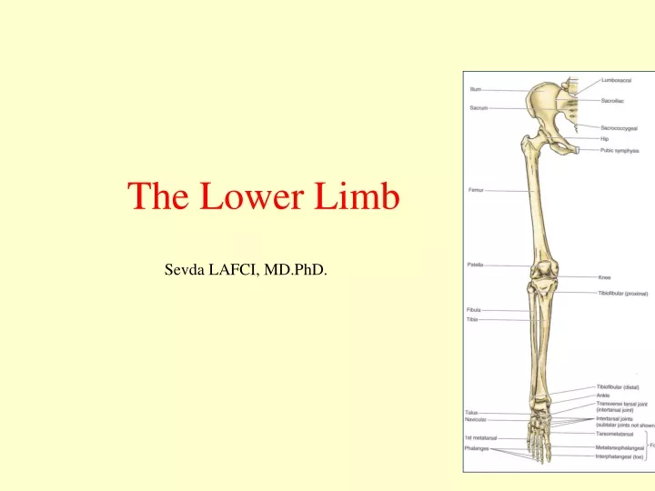
The Lower Limb. Sevda LAFCI, MD.PhD. The Lower Limb. The bones of the lower limb form the inferior part of the appendicular skeleton the organ of locomotion for bearing the weight of body stronger and heavier than the upper limb for maintaining equilibrium. The Lower Limb. 4 parts:

E N D
The Lower Limb Sevda LAFCI, MD.PhD.
The Lower Limb • The bones of the lower limb form the inferior part of the appendicular skeleton • the organ of locomotion • for bearing the weight of body • stronger and heavier than the upper limb • for maintaining equilibrium
The Lower Limb • 4 parts: • The pelvic girdle (coxae) • The thigh • The leg (crus) • The foot (pes)
The Lower Limb • Thepelvicgirdle: • formedbythehipbones (innominatebones-ossacoxae) • connectiontheskeleton of thelowerlimbtothevertebralcolumn
The Lower Limb • Thethigh • thefemur • connectingthehipandknee
The Lower Limb • Theleg • thetibiaandfibula • connectingthekneeandankle
The Lower Limb • The foot • distal part of the ankle • the tarsal bones, metatarsal bones, phalanges
The Lower Limb • 4 parts: • Thepelvicgirdle • Thethigh • Theleg • Thefoot
The pelvic girdleHip • the area from the iliac crest to the thigh • the region between the iliac crest and the greater torachanter of the femur • formed by the innominate bones-ossa coxae
The hip boneos coxae • large and irregular shaped • consists of three bones in childhood: • ilium • ischium • pubis • fuse at 15-17 years • joined in adult
The hip bonethe ilium • forms the superior 2/3 of the hip bone • has ala (wing), is fan-shaped • its body representing the handle • iliac crest: superior margin of ilium
The hip bonethe ilium • iliac crest • internal lip (labium internum) • external lips (labium externum)
The hip bonethe ilium • iliac crest end posteriorly “posterior superior iliac spine” at the level of the fourth lumbar vertebra bilat.* • iliac crest end anteriorly “anterior superior iliac spine • easily felt • visible if you are not fatty • *: it is important for lumbar puncture
The hip bonethe ilium • iliac crest can always be determined • Bilateral posterior superior iliac spina lies beneath a skin dimple* • The line connecting the right-l eft skin dimple is at the level of 2. sacral vertebra and the middle of the sacroiliac joint. • *: skin dimples formed by the skin and underlying fascia attached to the PSIS
The hip bonethe ilium • Tubercle of the crest is located 5cm posterior to the anterior superior iliac spine • ant. inf. iliac spine • post. inf. iliac spine difficult to identfy by palpation
The hip bonethe ilium • greater sciatic notch
The hip bonethe ilium • At the medial side • auricular surface for the sacroiliac joint
The hip bonethe ischium • it forms the posteroinferior part of hip • L-shaped • which passes inferiorly from the acetabulum • turns anteriorly to join the pubis • body • ramus
The hip bonethe ischium • at the inferior end of the body • ischial tuberosity • is covered by gluteus maximus muscle when the thigh is extended
The hip bonethe ischium • at the posterior part of the ischium • ischial spine (spina ischiadica) • separates the • greater sciatic notch sup. • lesser sciatic notch inf. G L
The hip bonethe ischium • the greater sciatic notch • is converted “greater sciatic foramen” by the sacrospinous ligament • pass the • the priformis muscle • the vessels and nerves of gluteal region G
The hip bonethe ischium • The lesser sciatic notch • is converted “lesser sciatic foramen” by the sacrospinous and sacrotuberous ligament • contains: • obtrator internus muscle • pudendal nerve • internal pudendal vessels L
The hip bonethe ischium • ramus • extends medially from the body • joins the inf. ramus of the pubis • form “ischiopubic ramus” which completes the obturator foramen
The hip bonethe pubis • forms anterior part of the hip bone • body, lies medially, joins body of the other ones • it’s called symphysis pubis (cartilaginous joint) • ramus (2) • superior ramus passes superiolaterally to the acetabulum • inferior ramus passes posteriorly, inferiorly, laterally to join ramus of ischium to form half of the pubic arch (ischiopubic ramus)
The hip bonethe pubis • the anterior border of the body is thickened “pubic crest” • its lateral end, pubic tubercule* *: main pubic attachment for the inguinal ligament- bony landmark
The hip bonethe obturator foramen • oval aperture • surrounded by the bodies and rami of the pubis and ischium • it lies inferomedial to the acetabulum
The hip bonethe obturator foramen • is nearly closed by the obturator membrane
The hip bonethe acetabulum • cup shape cavity • articulates with the head of femur • it’s names from Roman vinegar cup, it is called acetabulum • Until puberty the ilium, ischium and pubis are united by a “Y” shaped hyaline cartilage • At 15-17 years these bones fuse to form the hip bone (cartilage is replaced by bone)
The Lower Limb • 4 parts: • Thepelvicgirdle • Thethigh • Theleg • Thefoot
The thighFemur • thigh bone is femur • longest • strongest • heaviest bone • articulates with acetabulum and tibia • Height ≈ 4xlength of femur
The thighFemur • body (shaft) • ends (extremities) Proximal end: • head • neck • greater trochanter • lesser trochanter posterior aspect medial aspect
The thighFemur • Distal end: • broadened • articulates with tibia and patella medial aspect anterior aspect
The thighFemur • Proximalend: • head • neck • greatertrochanter • lessertrochanter posterior aspect medial aspect
The thighFemur • Proximal end: • Head • forms about 2/3 of a sphere • to fit deeply into the acetabulum • sometimes palpable when the thigh is rotated laterally in thin male posterior aspect medial aspect
The thighFemur • Proximalend: • head • neck • greatertrochanter • lessertrochanter posterior aspect
The thighFemur • neck • between head and body • to meet the body neck runs inferolaterally with angle of 125˚ • limited laterally greater trochanter posterior aspect
The thighFemur • Intertrochanteric line • between greater and lesser trochanter (anteriorly) • is produced by the attachment of the iliofemoral ligament (massive lig.) anterior aspect
The thighFemur • Intertrochanteric crest • unites greater and lesser trochanter, posteriorly posterior aspect
The thighFemur • Proximalend: • head • neck • greatertrochanter • lessertrochanter posterior aspect
The thighFemur • greatertrochanter • is large, rectangularprojection fromthejunction of theneckandthe body. posterior aspect
The thighFemur • greatertrochanter • is insertionformuscle of glutealregion • themostlateralpoint of thehipregion posterior aspect
The thighFemur • greatertrochanter • can be easilypalpated on thelateralside of thethigh • themostlateralpoint of thehipregion
The thighFemur • Proximalend: • head • neck • greater trochanter • lessertrochanter posterior aspect
The thighFemur • lesser trochanter • is located in the posteromedial surface • at the inferior end of the intertorachanteric crest • in the angle between the neck and body of the femur posterior aspect
The thighFemur • Body (shaft) • Linea aspera • in the middle of its posteriorly • has medial and lateral lips • Diverge inferiorly to form the supracondylar lines • not palpable, covered with large muscle
The thighFemur Body (shaft) • Pectineal line • runs from the lesser torachanter to the medial lip • tendon of the pectineal muscle inserts into it
The thighFemur • Distal end:
The thighFemur • Distal end: • Condyle, epicondyle • intercondylar notch • patellar surface • adductor tubercle
The thighFemur • Distal end: • broadened for articulation with tibia • 2 large “condyle” project posteriorly • are subcutaneous • easily palpable • separated by a deep U-shaped “intercondylar notch”
The thighFemur • Distal end: • at the center of the each condyle is a prominent “epicondyle” • tibial and fibular collateral ligaments are attached to the epicondyles