Download
1 / 1
10 likes | 196 Views
Fluorescence detection of proteins bound to a single DNA. John S. Graham, John F. Marko Northwestern University Materials Research Science & Engineering Center DMR-0520513.

E N D
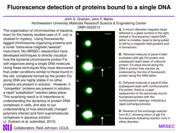
Fluorescence detection of proteins bound to a single DNA John S. Graham, John F. Marko Northwestern University Materials Research Science & Engineering Center DMR-0520513 A. 3-micron diameter magnetic bead tethered to a glass surface to the right, viewed in fluorescence; naked DNA tether is invisible, bead is being pulled to left by a magnetic field gradient and is fluorescent. B. Tethered molecule of panel A after incubation with gfp-Fis protein and subsequent wash away of unbound protein; Fis stays bound along the DNA in protein-free solution as evidenced by the line of fluorescent protein along the DNA tether. C. Tethered molecule of panel B after subsequent wash with nonfluorescent Fis protein; there is a rapid replacement of the previously bound fluorescent protein with the nonfluorescent species, indicating a rapid exchange process. D. Reverse of the exchange reaction from B-C, showing return of gfp-Fis fluorescence indicating reaction runs in either direction. The organization of chromosomes of bacteria, even for the heavily studied case of E. coli, is cloaked in mystery. Using fluorescently-tagged chromosome-organizing proteins and a novel “transverse magnetic tweezer” instrument, NU-MRSEC researchers have developed techniques to directly visualize how the bacterial chromosome protein Fis self-organizes along a single DNA molecule. Using these techniques they have discovered that under conditions similar to those found in the cell, complexes formed by the protein Fis along DNA are highly stable if no other proteins are present in solution. However, if “competitor” proteins are present in solution, a rapid “substitution” reaction takes place. This surprising result is of importance to understanding the dynamics of protein-DNA complexes in cells, and also to our understanding of how electrically charged polymers interact and form supramolecular complexes in aqueous solution. (J. Graham et al, submitted, 2010). Collaborators: Reid Johnson, UCLA.