Download
1 / 19
250 likes | 709 Views
Anatomy and Physiology of the Heart. Anatomy & Physiology/Cardiovascular System. Size and Location of the Heart. About the size of a an adult fist Hollow and cone shaped Weighs less than a pound Sits atop the diaphragm and is centered between the left and right lung.

E N D
Anatomy and Physiology of the Heart Anatomy & Physiology/Cardiovascular System
Size and Location of the Heart • About the size of a an adult fist • Hollow and cone shaped • Weighs less than a pound • Sits atop the diaphragm and is centered between the left and right lung. • The pointed apex is directed towards the left hip, while the base is directed towards the right shoulder.
Anatomy of the Heart • The heart is composed of four chambers: • The right and left atria, which make up the top portion of the heart, are receiving chambers. • The right and left ventricles, which make up the bottom portion of the heart, are the discharging and pumping chambers. • The lengthwise membrane that divides the heart is referred to as the interatrial septum OR interventricular septum, depending on which chambers it is dividing.
The Heart as a Pump • The heart functions as a double pump. • Pump #1 controls pulmonary circulation, which brings blood to and from the lungs. • Pump #2 controls systemic circulation, which carries blood throughout the body and back to the heart.
Veins and Arteries of the Heart • Superior and Inferior Vena Cava = located above and below the right atrium, carry deoxygenated blood from the body into the right atrium of the heart. • Pulmonary Arteries = Exit from the right ventricle and carry deoxygenated blood from the heart to the lungs.
Veins and Arteries of the Heart • Pulmonary Veins = Exit the lungs and carry oxygenated blood to the left atrium of the heart. • Aorta (Artery) = Exits the left ventricle of the heart and carries oxygenated blood to the body tissues.
Veins and Arteries of the Heart • Coronary Arteries = branch out from the aorta and deliver oxygenated blood to the heart tissues. • Cardiac Veins = carry deoxygenated blood from the heart tissues to the right atrium.
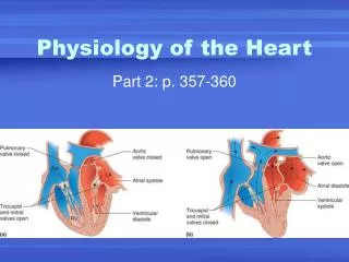
Valves of the Heart • The heart has four valves, which allow blood to flow in only one direction through the heart: • Atrioventricular (AV) Valves = located between the atrial and ventricular chambers on each side. These valves prevent backflow into the atria when the ventricles contract. • The left atrioventricular valve is also called the bicuspid valve because it is made of two flaps. • The right atrioventricular valve is also called the tricuspid valve because it is made of three flaps.
Valves of the Heart • The heart has four valves, which allow blood to flow in only one direction through the heart: • Semilunar Valves = located between the ventricles and the large arteries that exit them. These valves prevent backflow into the ventricles when the ventricles relax. • Pulmonary semilunar valves = located between right ventricle and pulmonary arteries. • Aortic semilunar valves = located between left ventricle and aorta.
Systole and Diastole • Systole = heart contraction • Diastole = heart relaxation • These terms refer to the contraction and relaxation of the ventricles, unless otherwise noted. • Blood Pressure readings are recorded as systolic pressure over diastolic pressure, which refer to the pressure in the arteries during contraction and relaxation, respectively. • A typical blood pressure reading is around 120/80 mmHg
Physiology of the Heart • Unlike skeletal muscle, cardiac muscle can contract independently, even if it is severed from all nerve connections. • Two systems act to regulate heart activity: • The nerves of the nervous system act to decrease and increase heart rate in response to environmental changes. • The intrinsic conduction system (nodal system) built into the cardiac muscle tissue.
Physiology of the Heart • There are four major parts that make up the intrinsic conduction system: • Sinoatrial (SA) Node = tissue located on the right atrium which starts each heartbeat and sets the pace for the whole heart. Also known as the pacemaker.
Atrioventricular (AV) Node = tissue located at the junction of the atria and ventricles. • Atrioventricular (AV) Bundle = groups of fibers located on the interventricular spetum. • Purkinje Fibers = spread throughout the muscle of the ventricle walls.
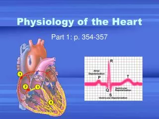
Physiology of the Heart • The electrochemical impulse that travels through the heart and generates each heart beat follows this path: • The impulse is generated in the SA Node • The impulse travels through the right and left atrium, triggering contraction of the atria. • The impulse reaches the AV Node where it is delayed long enough for the atria to complete contraction. • The impulse then travels along the Bundle of His to the Purkinje fibers and begins contraction of the ventricles at the apex of the heart.
Electrocardiography (EKG) • The impulses generated by the heart can be detected on the surface of the body. • A typical EKG includes three waves: • P Wave = the first wave, which signals the depolarization of the atria • QRS Complex = signals the depolarization of the ventricles • T Wave = the second wave, signals the repolarization of the ventricles.