Download
1 / 148
1.52k likes | 1.76k Views
Metabolic Systems. Energy requirement 能量需求. In order to maintain essential life processes To transform the chemical energy in the environment into electrical, mechanical, osmotic, and other forms of chemical energy . Digestion is a sequence of events that result in ever

E N D
Metabolic Systems Energy requirement 能量需求 • In order to maintain essential life processes • To transform the chemical energy in the environment • into electrical, mechanical, osmotic, and other forms of • chemical energy. • Digestion is a sequence of events that result in ever • finer degradation of the foodstuffs until it is broken • into monomersthat are absorbed into the body and • reassembled as needed into host structures.
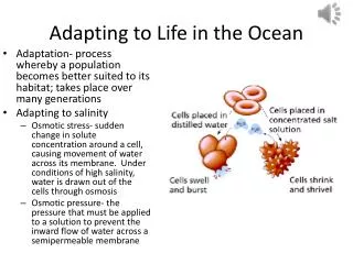
Metabolic Systems Dietary Requirements 營養需求 • Insects require basically the same nutrients as most • other animals: a source of carbon , essential amino • acids , essential fatty acids , inorganic salts , vitamins • and a source of sterol. Water is also an essential • nutrient. • Essential Nutrients are nutrients that require a diet • source since they cannot be synthesized from other • dietary nutrients or metabolic precursors.
The process of hydrolyticallybreaking large • macromolecules into their component subunits • suitable for absorption into cells. • The sources of transformed energy include the • ingested food that contains complex carbohydrates, • fats, and proteins. • Foods broken down in the alimentary tract to the • simpler components and absorbed through the wall • of the midgut into the hemolymph.
The circulatory system then transports these components • to all the cells of the body, which break them down even • further and capture the chemical energy they contain. • Each cell may use the components immediately or they • may be used to synthesize reserves for later use. • These processes of food breakdown, utilization, and • storage are strikingly similar in almost living things. • Insects show diverse morphologies of their digestive • systems because of their diverse diets. Generally, the • higher the protein content, the shorter the intestine; the • lower the protein content, the longer the intestine.
The metabolic systems of only a small number of insect • species have been examined, most often cockroaches, • blow flies, fruit flies, or caterpillars. • The evidence for the existence of complete metabolic • pathways in insects has often been based on the • presence of certain key enzymes, reaction end products, • or intermediates. • The determination of metabolic pathways in insects is • complicated by the presence of symbiotic • microorganisms that may provide some of the steps • missing in the insect. • This symbiotic contribution makes it difficult to • establish whether the metabolic pathways are actually • present in the insect system.
Energy production from food Chemical energy transformation Ingestion Digestion: Digestive enzymes Symbiotic microorganism: vitamins etc Absorption:: Utilization Storage Cockroach, blow fly, fruit fly, caterpillars
The Insect Alimentary Tract Foregut(stomedeum)前腸– Ectodermal Cuticle; intima; shed and renewed 。Food fragmentation; Food storage. Midgut(mesenteron)中腸– Endodermal 。Food Digestion; Food absorption Peritrophic membrane Hindgut(protodeum)後腸–Ectodermal 。Collects and conducts waste products for excretion 。Nutrient reabsorption 。 Water and Salt balance and osmotic regulation
Diversity in insect feeding and the alimentary structures (no typical digestive tract among insects ) Diversity in diets Mouthpart structures Specialization of digestive tract Ecological niches Discontinuous feeders Predatory or carnivorous species Food storage mechanism Continuous feeders Phytophagous insects
General Structure of Alimentary Canal The digestive tract consists of a tube of epithelial cells running from the mouth to the anus.
Three region of insect alimentary canal Foregut: stomodeum; ectoderm; cuticle Midgut: endoderm; peritrophic membrane Hindgut: proctodeum; ectoderm; cuticle
Foregut, Midgut, Hindgut • The stomodeum and • proctodeum both • arise as invaginations • of the embryonic • ectoderm and produce • the foregut and hindgut. • The midgut forms from • endodermal tissues and • connects with the • foregut and hindgut • during embryogenesis.
Anterior structures and the foregut Evolution of insects from a primitive annelid ancestor
Anterior structures and the foregut Mouthparts (mandibulate insects)口 Biting; Cutting; Grinding Preoral cavity Cibarium; Hypopharynx ; Salivarium Stylet sheath: plant-feeding hemipterans Preoral Cavity (cibarium)前口腔 Enclosed by the mouthparts and opens into the oral cavity or mouth Haustellate口吻(proboscis) Cibarium 食料腔
Extrinsic visceral muscles Intrinsic visceral muscles Longitudinal muscles Circular muscles (labium)
Salivary gland唾腺 for salivary secretions Labial Glands下唇腺 Evolved from the epidermal cells of labium Salivary duct Saliva: solvent for food; lubricates the mouthparts Digestive enzyme: amylase, invertase, protease, chitinases Predatory insects: inject saliva into their hosts Toxin: act on nervous system due to host paralyze Anticoagulants: blood feeding insects
Salivary gland唾腺 Salivary enzymes may perform exodigestion: Pectinase (aphids) Hyaluronidase Blood feeding insects (blood feeders) Anticoagulins: agents to increase the rate of blood flow Predatory insects: inject saliva into their hosts Lipolytic and proteolytic enzymes in assassin bug. Silk-producing lepidopterans: labial gland as silk gland salivary gland evolved from mandibular gland
Pharynx咽- the first region of foregut characterized by dilator muscles from the ventral tentorium and the dorsal fronts muscular sucking insects: vacuum lumen (pump) Esophagus食道– Simple tubular undifferentiated part, serves to pass food from the pharynx to the crop. Diverticula分枝盲囊: Adult Diptera and Lepidoptera : Pharyngeal receptors can determine and separate the ingested food
Crop嗉囊- Enlarged area for food storage. Food storage May be folded, or modified into a lateral diverticulum (分枝 盲囊)in fluid feeders. The secretion and absorption do not occur in the crop. Because the intima limitation, but crop of Periplaneta to be permeable to free fatty acid. Digestion Digestion can occur because salivary enzymes pass back with the food and midgut enzymes are regurgitated forward. The proventricular valve prevents the movement of solid food, but not the regurgitation of fluids.
Proventriculus前胃 Generally a grinding function (muscular structure); various Modified control the passage of food; retaining food (valve). Muscular spincter: beetles Gizzard lined with teeth: cockroach Backwardly pointing spines: fleas The proventriculus projects into the crop and armed with spines: bees
Cockroaches and crickets have sclerotized plates or teeth (denticles) for breaking up food Denticles absent in fluid feeders Fleas have spines for rupturing RBC
Bees have denticles (mobile lips) arranged as sieve-like spines to strain pollen from the nectar. The pollen is collected as a bolus and passed to the midgut for digestion while the nectar is retained in the crop for regurgitation to be formed into honey, back at the hive (honey = bee barf!).
Midgut 中腸 Digestion and absorption Most distinctive midgut cells are tall and columnar with regular microvilli forming a brush-border on the lumen side. The microvilli supported by abundant of actin filaments, great increase the area of the cell membrane for nutrients absorption. Schistocerca americana: 9000 microvilli/cell about 500cm2 total midgut lumen area.
Cell types of midgut Columnar cells: principle cells; Regenerative cells: group in nidi Goblet cells: single cell distribution Endocrine cells: single cell distribution Gastric caecae:
Columnar cells 柱狀細胞 Principle cells Endodermal origin, microvilli and folds (abundant) Digestive enzymes synthesis and secretion Nutrients absorption
These columnar cells contain abundant mitochondria, ER, Golgi bodiesand serve to both secrete digestive enzymes and absorb the products of digestion. The digestive enzyme secreted byexocytosis or apocrine secretion. The columnar cells have tight junctions and septate desmosome and are covered by a basement membrane on the hemolymph side
Regenerative cells再生細胞 Columnar cell regenerate Midgut cells regenerate at the rate of 40-120 hours
group in nidi The midgut is surrounded by poorly developed layers of muscles over the epidermal cells. In the converse of the foregut, the circular muscles cover the columnar cells with the longitudinal muscles on the hemolymph side. The muscle layers are bounded by a thin connective tissue sheath.
Goblet cells杯狀細胞 single cell distribution Regulate the ion transport: potassium ion Processing of K+ transport: [V-ATPase → H+ pump] [K+/H+ antiporter] [ it’s high energy consumption (10% of total ATP)]
Goblet cells occur in the midgut of Lepidoptera and Trichoptera. They secrete K+ from the hemolymph into the lumen of the intestine. They are especially important in Lepidoptera larvae where the midgut is alkaline. This is important because both the toxin of Bacillus thuringiensis and baculovirus are active in lepidopteran larvae because of the alkalinity (pH 10) of the midgut. Goblet cells may also assist in excreting excess K+ from the hemolymph. They may also participate in storage excretion of metals. Deposit excretion and discharge during moult. Rhodinus: hemoglobin→hematin
Endocrine cells內泌細胞 single cell distribution Involved in the regulation of enzyme production Insulin family: glucagons, somatostatin, β-endorphins Tachykinin family: myotropins: as cardioaccelerators stimulate muscle contraction FMRF Famide-immunoreactive peptides function in digestion Allatostatin-like peptides: regulate CA activity
Gastric caecae胃盲囊: Increase the surface area for secretion and absorption Create a countercurrent flow within the gut for efficient of digestion and absorption Food detoxification: secondary compounds
Peritrophic Membrane圍食膜 Peritrophic membrane: peritrophic matrix (PM) Consists of chitin microfibrils, proteins, carbohydrates PM consist of chitin and proteins, is secreted by the midgut cells, function as a wall to separate the food from midgut epithelium, and they does not invaginate into the cecae. Composition - chitin and protein: 3.7-12.9% chitin; 21–47 % protein. Insects secrete no mucous and the PM is chitin which is composed of acetylglucosamine -a mucopolysaccharide. Biochemically mucopolysaccha-rides and mucoprotein and chito-protein are closely related.
Functions of Peritrophic membran Protection Selective permeability Digestion area: enzymes binding Detoxification Barrier of microorganism
Protection PM is present in most insects whether they feed on solid food or only fluids. For prevent the food particle contact withthe epithelial cells of midgut, avoiding damage. The PM is absent in Homoptera and Heteroptera that feed only on plant juices. Harmful chemical, like tannic acid, may be keep at the small pockets which formed by PM, and then eliminate with feces. These phytophagous insects must have certain physicochemical properties of the matrix in the pockets.
Barrier The PM likely acts as a barrier to micro-organisms thus reducing infection by protecting the gut epithelium from abrasions by the food and as a barrier to bacteria. Separation Efficient for enzyme activity. Digestive enzyme binding Aminopeptidase
Ultrafilter PM pores are up to 0.2μm across and act as an ultrafilter to screen out large molecules. No hindrance to digestion products or digestive enzymes. P.M. permeable both ways to H2O, salts, acids, mono- and disaccharides and amino acids. High M.W. sugars (starch) and proteins (casein, albumin, gelatin) are retained. Diptera: 4-5 nm Grashopper: 25-35nm
Types of PM formation Type I: PM secret from whole cells of midgut Orthoptera, Odonata, Coleoptera, Hymenoptera Ephemeroptera, Lepidoptera larvae This type is a delamination from the whole surface of the midgut. Consists of concentric lamellae - separate thin sheets elaborated from the cell surface - a chitin containing material. Wasp and bee: 1/2 doz. membrane/day several layers, one inside the other. In Periplaneta, the PM consists of three layers of fibrils deposited at 60º to 90º to each other. The fibrils are formed into a network around the microvilli of the epithelium.
Type II: anterior region cells of the midgut Diptera, Dermaptera Diptera and some other orders, the PM is secreted as a viscous fluid at the anterior end of the midgut. This fluid is forced through a mold press from by the stomodeal invagination and the wall of the midgut so that its forms a tube which becomes the membrane. This membrane is formed continuously at a rate of 6 mm/h in Eristalis (Diptera). Usually larmina form is present.
Countercurrent flow creation Endoperitrophic space Ectoperitrophic space Gastric caecae
Digestion of cockroach Preliminary digestion: crop Further digestion: midgut: slight acidic to neutral in pH Fermentation chamber locate at anterior hindgut: alkaline in pH; Bacteria symbionts Water reabsorption occur at rectum Dry fecal pellet form at rectum and then deposit The digestive process about 20 hour for solid food
Digestion of plant feeder Specialization of alimentary tract Digestion of the larvae of Lepidoptera insects No digestion occur in foregut Digestion taking place in endoperitrophic and ectoperitrophic space Countercurrent flow mechanism; High pH value in midgut Digestion of liquid plant feeding: Homoptera, Hemiptera Dilute nutrients; elongation of alimentary tract Filter chamber specilization
Digestion of proteins Protein digestion: proteolytic enzyme: proteases; peptidases Endopeptidases: cleave internal peptide bonds Exopeptideses: remove terminal amino acids from peptide chain Dipeptidase - splits free dipeptides amino acids. Special Proteases Keratinase - digests wool. Tineola, dermestids, Mallophaga Tineola: Cysteine desulfhydrase Collagenase - splits connective tissue component of animals, Lypoderma and Lucilia綠蠅
Endopeptidases: cleave internal peptide bonds Serine proteases: serine Trypsin: cleave protein chains on the carboxyl side of basic a.a. Trypsin breaks the peptide bond at lysine or arginine (dibasic amino acids). Break down protein to peptones and polypeptides (absorb form). Chymotrypsin: cleaves protein chains on the carboxyl side of aromatic a.a. tyrosine, phenylalanine, tryptophan
Exopeptideses: remove terminal amino acids from peptide chain Carboxypeptidases: carboxyl end of peptide chain Aminopeptidases: N-terminal of the peptide chain, Metal ion as co-enzyme