Download
1 / 71
710 likes | 828 Views
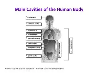
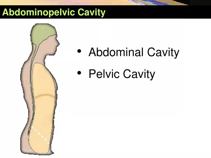
Abdominopelvic Cavity. Abdominal Cavity Pelvic Cavity. Landmarks of abdomen. Xiphoid process Costal margin Tip of the ninth costal cartilage Iliac crest Anterior superior iliac spine Pubic tubercule. Pubic crest Pubic symphysis. Boundaries of the A bdomen. Boundaries

E N D
Abdominopelvic Cavity • Abdominal Cavity • Pelvic Cavity
Landmarks of abdomen • Xiphoid process • Costal margin • Tip of the ninth costal cartilage • Iliac crest • Anterior superior iliac spine • Pubic tubercule. • Pubic crest • Pubic symphysis.
Boundaries of the Abdomen • Boundaries • Superiorly: Xiphoid process, lower border of costal arch, 11th and 12th ribs, vertebra T12 • Inferiorly: Superior border of pubic symphysis, pubic crest, pubic tubercle, fold of inguinal canal, anterior superior iliac spine, iliac crest, spinous process of L5
Landmarks of abdomen 1. Xiphoid process. 2. Costal margin. 3. Tip of the ninth costal cartilage. 4. Tendinous intersections. 5. Umbilicus. 6. Iliac crest. 7. Anterior superior iliac spine. 8. Linea semilunaris. 9. Linea alba. 10. Inguinal ligament. 11. Pubic tubercule. 12. Pubic crest. 13. Pubic symphysis.
Landmarks of abdomen • Horizontalplanes • Transpyloricplane (L1): Halfwaybetween the jugularnotchand the symphysispubis • Subcostalplane (L3): connects the lowestpoints of the twocostalarches. • Supracristalplane (L4): joins the the highestpoints of the twoiliaccrests. • Intertubercularplane (L5): connects the tubercles of the rightandleftiliaccrests
Landmarks of abdomen • Sagittalplanes • Medianplane: coincideswith the lineaalbaandpassesthrough the umbilicus. • Lateralplanes (midclavicularlines): connectsmidpoint of eachclavicletomidpoint of inguinalfold.
Landmarks of abdomen • Horizontalplanes • Transpyloricplane (L1): Halfwaybetween the jugularnotchand the symphysispubis • Subcostalplane (L3): connects the lowestpoints of the twocostalarches. • Supracristalplane (L4): joins the the highestpoints of the twoiliaccrests. • Intertubercularplane (L5): connects the tubercles of the rightandleftiliaccrests • Sagittalplanes • Medianplane: coincideswith the lineaalbaandpassesthrough the umbilicus. • Lateralplanes (midclavicularlines): connectsmidpoint of eachclavicletomidpoint of inguinalfold.
Landmarks of abdomen • Horizontalplanes • Transpyloricplane (L1): Halfwaybetween the jugularnotchand the symphysispubis • Subcostalplane (L3): connects the lowestpoints of the twocostalarches. • Supracristalplane (L4): joins the the highestpoints of the twoiliaccrests. • Intertubercularplane (L5): connects the tubercles of the rightandleftiliaccrests • Sagittalplanes • Medianplane: coincideswith the lineaalbaandpassesthrough the umbilicus. • Lateralplanes (midclavicularlines): connectsmidpoint of eachclavicletomidpoint • of inguinalfold.
Auricula = Kulak Kepçesi The abdominal regions - Four quadrants • Medianplane • Horizontalplane(supracristalplane) • Left and right upper quadrants • Left and right lower quadrants
The abdominal regions - Nine regions Epigastric region R. hypochondriac region L. hypochondriac region R. lateral regions L. lateral regions R. inguinal region Umbilical region L. inguinal region Pubic region
The Layers of Anterolateral abdominal wall Layers(from superficial to deep) • Skin • Superficial fascia • Anterolateral muscles • Transverse fascia • Extraperitoneal fascia • Parietal peritoneum
The Layers of Anterolateral abdominal wall Layers(from superficial to deep) • Skin • Superficial fascia • Anterolateral muscles • Transverse fascia • Extraperitoneal fascia • Parietal peritoneum
The Layers of Anterolateral abdominal wall Anterolateral abdominal wall Posterior abdominal wall
The Layers of Anterolateral abdominal wall • Skin • Superficial fascia • Deep fascia • Muscles • Transversalis fascia • Extraperitoneal fascia • Peritoneum
Superficial Facia Divisions below umbilicus: • Fatty layer (Camper’s fascia) continuous with the superficial fascia over the rest of the body. • Membranous layer (Scarpa’s fascia)passes over the inguinal ligament to fuse the deep fascia of the thigh (fascia lata) approximately one fingerbreadth below the inguinal ligament. In the midline, it is not attached to the pubis but instead forms a tubular sheath for the penis (clitoris). In the perineum, it attaches on each side to the margins of the pubic arch and is know as Colles’ fascia.
Superficial Facia • Firmly anchored in the dermis, but adhereonlylooselyto the deep facia of EO. • Has twolayers: • Superficialfattylayer (Camper’s fascia) • Deepmembranouslayer (Scarpa's fascia)
Superficial Facia • Superficialfattylayer (Camper’s fascia) • In the male, the fatdisappearsover the penis. • In the female, fattylayercontinuesinto the labiamajora.
Superficial Facia • Deepmembranouslayer (Scarpa's fascia) • Extendsonto the dorsum of penis, known as fundiformligament. • Formssuperficialpenile facia • Surrounds the scrotum (dartosfascia). • Inbothsexes, bothlayersbecome the superficial facia of the perineum.
Superficial Arteries • Lateral • Anteriorintercostal a. • Subcostal a. • Lumbar a. • Median • Superior epigastric a. • Inferior epigastric a. • Inferior • Superficial epigastric a. • Superficial iliac a.
SuperficialVeins lateral thoracic subclavian thoracoepigastric portal paraumbilical S. epigastric femoral S. circumflex iliac
Caput Medusae(Medusa Head) SuperficialVeins
Lymph Drainge • Superficial lymph vessels above the level of the umbilicusdrain upward into the pectoral Ln. • The vessels below this level drain downward into the superficial inguinal Ln.
Abdominal Muscles Anterior Group Lateral Group • External Oblique • Internal Oblique • Transversus • Rectus Abdominis • Pyramidalis
External Oblique Abdominis • General direction of fibers: downward, forward and medially (run down and inward) • Thelargestabdominalmuscle. • Margins: • Posteriormarginis fleshyandvertical. • Inferiormarginis entirelytendinous. • Thickensbetweentheanteriorsuperioriliacspineandthepubictubercleandformstheinguinalligament.
External Oblique Abdominis • Structures • Inguinal ligament • Lacunar ligament • Canalisinguinalis • Superficial inguinal ring • Triangular-shaped defect in aponeurosis of obliquusexternusabdominis above pubic tubercle • Deepinguinal ring
External Oblique Abdominis • Tendonfibers in theupperportion of theaponeurosisdecussate in thelineaalba. • Compressesthe abdomen.
Internal Oblique Abdominis • Deep to obliquusexternusabdominis • General direction of fibres: upwards, forwards and medially • Does not have a freeposteriorborder. • Has freeinferiorborderpassingovertheinguinalligamentandcontributingtheformation of conjointtendon.
Internal Oblique Abdominis • Superiorly, its aponeurosis splits into an anterior and a posterior layer along the lateral edge of the rectus. • These layers constitute the rectus sheath and meet their fellows of the opposite side in the linea alba. • Arcuateline
Transversus Abdominis • Deep to obliquusinternus • General direction of fibers: run horizontally forward.
Transversus Abdominis • Inguinal falx(conjointtendon) • Obliquusinternusabdominis has a lower, free border that arches over spermatic cord • Inserted with transversusabdominis fiber into medial part of pecten of pubis • Cremastermuscle • Dirived from the lower fibers of the obliquusinternusabdominis and transversusabdominis • Around the spermatic cord and testis