Download
1 / 30
310 likes | 624 Views
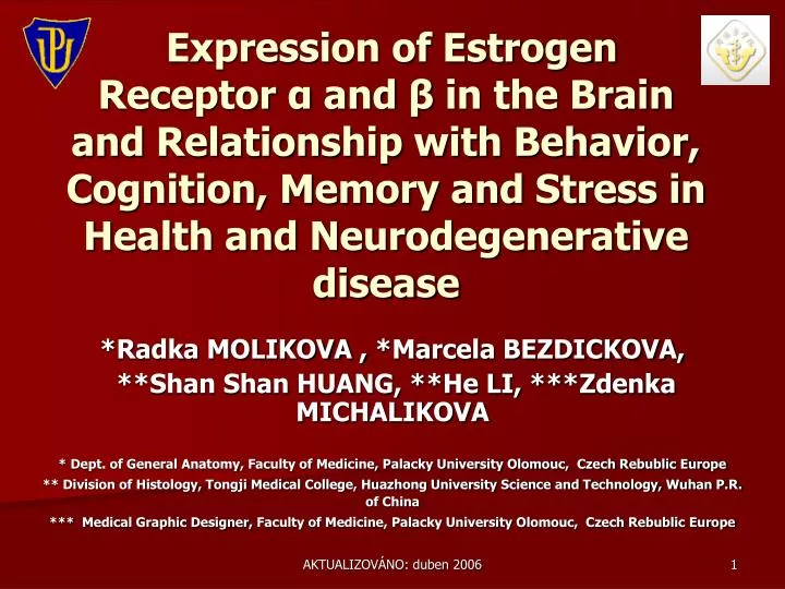
Expression of Estrogen Receptor α and β in the Brain and Relationship with Behavior, C ognition , Memory and Stress in Health and Neurodegenerative disease. *Radka MOLIKOVA , *Marcela BEZDICKOVA, **Shan Shan HUANG, **He LI, ** *Zdenka MICHALIKOVA

E N D
Expression of Estrogen Receptor αand βin the Brain and Relationship with Behavior, Cognition, Memory and Stress in Health and Neurodegenerative disease *Radka MOLIKOVA , *Marcela BEZDICKOVA, **Shan Shan HUANG, **He LI, ***Zdenka MICHALIKOVA * Dept. of General Anatomy, Faculty of Medicine, Palacky University Olomouc, Czech Rebublic Europe ** Division of Histology, Tongji Medical College, Huazhong University Science and Technology, Wuhan P.R. of China *** Medical Graphic Designer, Faculty of Medicine, Palacky University Olomouc, Czech Rebublic Europe AKTUALIZOVÁNO: duben 2006
Introduction • ERαand ERβ are an important form of steroid hormones with multiple targets, in the body and brain, and exerting ubiquitous effects on behavior. • In the brain these receptors are regionally specific, but both have widespread distributions, which are largely non-overlapping[1, 7, 8]. • In the CNS, estrogens are well known to cause a importance for control of REPRODUCTION, including SEXUAL BEHAVIOR but not only in the context of reproduction, but also with reference to COGNITION AND MEMORY[1, 4, 6].
Purpose • The physiological function of ERαand ERβis under intense study but certain results indicate that ERαand ERβhavedifferent or event opposite biological actions[1]. • Hoon´s preliminary studies show that hormone receptors, estrogen receptors (ERs), and cAMP response elements binding protein (CREB) are present in the mitochondrial matrix of neurons and ER and CREB could be regulated by novel signaling pathways in the intact central nervous system and that their functions in the mitochondria might be important in neuronal survival[10].
On the basis of newly emerging complexities of estrogen's mechanisms of action it is important[1, 7]: • To distinguish which pathways of ERαand ERβ are involved in modifying which behavior, cognition and memory. • To study the mechanisms by which estrogens mediate behavior, cognition and memory in which parts of the brain during increasing (pEGFP-C2- ERα,β) and decreasing (siRNA) expression of ERαand ERβ. • To compare mediate mechanism and biological function of ERαand ERβ in the brain and potential relation to neurodegenerative diseases (PD, AD, HD).
ERαand ERβ • ERαis localized on human chromosome 6, in contrast to ERβ, which sits on chromosome 14.ERαand ERβ thus represent two separate gene products and share relationship[1, 6]. • ERαis highly expressed in the classical estrogen target tissues such as uterus, mammary gland, bone and cardiovascular systems, whereas ERβis mainly expressed in non-classical tissues such as prostate, ovary and urinary track[2]. • Overall distribution of ERαand ERβ in different tissues figure 1[1].
Gustafsson A.J. 1999 Estrogen receptor β-a new dimension in estrogen mechanism of action. Journal of Endocrinology 163, 380. [1] Figure 1
ERαiswidely expressed in the hypothalamus. The distribution of ER αconcentrated in the band of Broca, the medial mamillary nucleus, the medial preoptic area, the paraventricular nucleus,and the ventromedial nucleus[3]. • ERαhas been localized in the hippocampus, with dorsal vs. ventral differences in the ERαexpression[5]. • Sensitive binding study have also showed the expression of estrogen receptors in cells of the neocortex, where their accurate localization and function is not so clear (resp.unknow), but may involve mediation of estrogen action on sexual behavior[4].
Neurodegenerative disease • Protein aggregation in the brain has been observed in the brains of a big sort of diseases characterized of progressive neurodegenetation, such as • Alzheimer Disease (AD) • Parkinson Disease (PD) • Huntington Disease (HD) • Creuzfeldt Jakob Disease (CJD) etc. • Abnormal protein aggregations are associated with gene mutation, protein posttranslational aberrant modification, chronical viral infection in the brain, brain aging and environmental toxin as described as follows.
ALZHEIMER DISEASE (AD) • AD is a progressive, degenerative disorders of uncertain causes, although abnormal metabolism and deposition of β-amyloid protein appears to be closely linked to pathogenesis. • AD is the most common cause of dementia. Its incidence rises from less than 1% per year to more than 7% per year, and its prevalence from 3% to almost 50%, between the ages of 65 and 85 years[13]. • This translates into a prevalence of 10-20mil. cases worldwide. Men and women are affected with equal frequency, when adjusted for age[13,14].
AD is defined by characteristic histopathologic features, especially neurofibrillary tangles and neuritic (senile) plaques[13,14]. • Examination of the brain will reveal variable cortical atrophy with widening of the cerebral sulci that is most pronounced in the frontal, temporal and parietal lobe. • The progressive loss of neurons in the entire cortex leads to slowly progressive mental deterioration. • Clinical symptoms include the loss of recent memory (the most common early sign) and loss of long-term memory. The disease is progressive and leads to immobility in about ten years[14].
[9]Johnson,K.A., Becker, J.A. The Whole Brain Atlas. Harvard Medical School.
PARKINSON DISEASE (PD) • Parkinsonism occurs in all ethic groups, in the United States and Western Europe it has a prevalence of 1-2/1000 population, with an approximately equal sex distribution. The disorder becomes increasingly common with advancing age. • It is characterized by expressionless face (masklike), tremor 4-6Hz („pill-rolling“), hypokinesia, rigidity, and abnormal gait and posture[13,14]. • It is important to compare HD with excessive movement to PD with decreased movement[13].
Etiology (multiple) • Idiopathic (most common) • Postencephalitis • Autoimmune dysfunction • Trauma • Drugs (illicit drug MPTP). • Location • Substantia nigra and other brainstem centers, cell loss in the globus pallidus and putamen and the presence of Lewy Body, containing α-synuclein in the basal ganglia, brainstem, spinal cord, and sympathetic ganglia. • Proces • Decreased dopamine in corpus striatum. • Treatment • Dopamine agonists.
HUNTINGTON DISEASE (HD) • HD is a hereditar disorders of the nervous system characterized by the gradual onset and subsequent of chorea and dementia. It occurs throughout the world and in all ethnic group. Its prevalence is about 5 per 100 000 population[13]. • HD is an autosomal dominant disorder. HD is caused by the increase in the length of a CAG triplet repeat present in a gene located on chromosome 4p16.3. The product of the mutated gene is an abnormal protein - HUNTINGTINE[9].
The typical onset of HD appears usually in the 4th decade (typically between 30 and 50 years of age), juvenile and late-onset cases are rare. • First symptoms are motor or mental changes (choreatic dyskinesias, later dystonic or rigid symptoms, personality and behavioral changes, affective disorders, progressive cognitive deficits and later dementia)[9,13]. • The pathophysiology of HD relates to degeneration of GABA neurons (typical inhibitory neurons) in the striatum. Decreasing functioning of inhibitory neurons leads to increased movements[14].
[9]Johnson,K.A., Becker, J.A. The Whole Brain Atlas. Harvard Medical School.
Material and Methods-The Present State Present State
Single Imunohistochemistry ER+ 3V Paxinos, G., Franklin, K.B.J. The Mouse Brain in Stereotaxic Coordination. [11] MUS MUSCULUS Bregma - 1,70mm Dilution 1:400 ER+
HYPOTHALAMIC AREA 3V VMHDM DM VMHVL 3V PeF LH MCLH magn. 10x magn. 20x LH 3V magn. 40x magn. 40x
CEREBRAL CORTEX AREA * S1Tr/S1BF magn.10 x S1Tr/S1BF magn.20 x * S1Tr/S1BF magn.40 x S1Tr/S1BF magn.100 x Expression of ER in the cerebral cortex area have been verify by RT PCR & Western Blot.
Acknowledgements • I would like to thank my supervisor Prof. LI He, PhD., for his expert advice and great help, Shen Shen Huangfor her devoted guidance relentless energy, enthusiasm, with which they guided me through the entire duration of my study in the Tongji Medical College. • Last but by no means the least; I must thank my collegues from Division of Histology, Tongji Medical College for their enormous tolerance and support. This project was supported by scholarship cooperation Ministry of Education Czech Republic and Ministry of Education P. R. of China in the Tongji Medical College in the year 2005/06.
References • Gustafsson, A.J. (1999) Estrogen receptor β– a new dimension in estrogen mechanism of action. Journal of Endocrinology 163, 379–383. • Liqin, Z., Tzu-wei, W., Roberta, D.B. (2004) Estrogen receptor subtypes alpha and beta contribute to neuroprotection and increased Bcl-2 expression in primary hippocampal neurons. Brain Research. Feb. 1010:22–34. • Pinzone,J.J et al. (2004) Molecular and Cellular Determinants of Estrogen Receptor ß Expression. Molecular and cellular biology, June, 24(11): 4605–4612. • Greenstein, B., Greeinstein, A. Color Atlas of Neuroscience: Neuroanatomy and Neurophysiology. Sttutgart: Thieme SRN, 2000. ISBN 0-086577-710-1. • Hart, S.A., Patton, J.D., Woolley, C.S. (2001) Quantitative analysis of ER alpha and GAD colocalization in the hippocampus of the adult female rat. J Comp Neurol. Nov 12;440(2):144-55. • Enmark, E., Pelto-Huikko, M., Grandien, K., Lagercrantz , S., Lagercrantz, J., Fried, G., Nordenskjold, M., Gustafsson, J.A. (1997) Human estrogen receptor ß – gene structure, chromosomal localization, expression pattern. Journal of Clinical Endocrinology and Metabolism 82:4258–4265. • Rissman, E.F., Wersinger, S.R., Fugger, H.N., Foster, T.C. (1999) Sex with knockout models: behavioral studies of estrogen receptor alpha. Brain Res. Jul 17;835(1):80-90. • Lundholm et al. (2004) Gene expression profiling identifies liver X receptor alpha as an estrogen-regulated gene in mouse adipose tissue.Journal of Molecular Endocrinology.32 (3):879-892. • Roth,J., Zidovska,J., Uhrova,T., Doubek,P. at al Huntington´s disease and ethical problems related to the diagnosis. Europ: Psychiatry 2000;suppl. 2:395–396. • Hoon,R. (2006) Estrogen Regulation of Mitochondrial Transcription in HD. Boston University Medical Campus, dept. Of neurology and National Institute of Neurological Disorders and Stroke at the link: http://researchresources.bumc.bu.edu/abstract/1R01NS052724-01.htm • Paxinos, G., Franklin, K.B.J. The Mouse Brain in Stereotaxic Coordination. The Prince of Wales Medical Research Institute, Australia and McGill University Montreal, Quebec, Canada. San Diego: Academic Press, USA, 2001. ISBN 0-12-547636-1. • Johnson,K.A., Becker, J.A. The Whole Brain Atlas. Harvard Medical School. At the link: www.med.harvard.edu/AANLIB/home.html • Brown,E. Basic Concepts in Pathology. Beijing: McGraw-Hill 2002 ISBN 7-81071-389-2 • Greenberg, D., Aminof, M., Simon, R. Clinical neurology. McGraw-Hill 5.th edition 2002 ISBN 7-117-4950-2
ESTROGEN RECEPTOR www.komsta.net www.breastcancer.org Cell with estrogen receptors, estrogen, and helper proteins.A Estrogen receptorB EstrogenC Estrogen helper proteinsD nucleusE DNA genetic material www.ks.uiuc.edu www.few.vu.nl
Protocol for cloning eukaryotic DNA fragments in lambda phage An example of DNA cloning using bacterial plasmids Formation of a recombinant DNA molecule
Department of AnatomyLeipzig University, Germany Molikova Radka Bezdickova Marcela
Thank you for your attention!