Download
1 / 41
410 likes | 521 Views
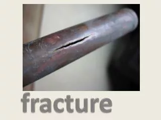
Fracture. Definition of Fracture. It is disruption of bone continuity. Although most of #s occur as a result of a single episode by a force powerful enough to fracture a normal bone, there are 2 types of # in which this is not so : Pathological fract. Stress fract.

E N D
Definition of Fracture • It is disruption of bone continuity. Although most of #s occur as a result of a single episode by a force powerful enough to fracture a normal bone, there are 2 types of # in which this is not so : • Pathological fract. • Stress fract.
Pathological fracture : It is one in which a bone is broken through an area weakened by pre-existing disease , & by a degree of force that would have left normal bone intact e.g osteoporosis , O.M. , bone tumours.
Stress fracture : Bone , like other materials , reacts to repeated loading . On occasion , it becomes fatigued & a crack develops e.g military installations , ballet dancers & athletes.
AO Classification • A : Simple fract. A simple bone fracture is a less damaging type of fractures. There is only bone damage • B : Wedge fract.A wedge fracture is a vertebral compression fracture occurring anteriorly or laterally. Viewed, the affected vertebra resembles a wedge • C : Complex fract. this type of fractured bone causes a large amount of damage to the soft tissue around the bone
Closed (This type indicates that there is no communication between the external surface of the body and the fracture) • Open (There is a communication between the fracture and the skin. This could occur because the displacement of the bone ends has caused one or both to pierce the skin, or because an external force has pierced the skin, soft tissues, and fractured the bone. This type of fracture is an additional cause for concern because of the possibility of infection
Fractures-Open Classification I <1cm long, minimal contamination, low energy force II >1cm long, moderate contamination and force III High energy, comminuted fx, extensive tissue damage, extensive contamination, arterial injury
Stable fractures • Occur when a piece of the periosteum is intact across the fracture • External or internal fixation has rendered the fragments stationary • Unstable fractures • Grossly displaced • Poor fixation
Mechanism of Injury Classification • Direct trauma • Tapping fractures • Crushing fractures • Penetrating fractures - High velocity missiles > 2500 f/s - Low velocity missiles < 2500 f/s
Indirect trauma • Traction or tension fract. • angulationfract. • Rotational fract. • Compression fract
Fracture Healing • Reparative process of self-healing (union) occurs in the following stages: • Fracture hematoma (d/t bleeding, edema) • Granulation tissue → osteoid(3 – 14 days post injury) • Callus formation (minerals deposited in • osteoid
Reparative process of self-healing (union) occurs in the following stages: • Ossification (3 wks – 6 mos) • Consolidation (distance between fragments decreases → closes). • Remodeling (union completed; remodels to original shape, strength)
Bone fracture healing and especially fracture healing times are common concerns. Bone fracture healing times vary considerably depending on: • the age and general health of the patient, • the bone affected - as well as the type of bone, • the type of fracture, and • other aspects of the injury and overall state and condition of the affected area e.g. presence of infection or other complications.
Diagnosis • Clinical picture • Radiography
Clinical Features of Fracture • History of trauma • History : - Trauma - Direct - Indirect • Pathological fractures Here the bone is weakened by pathological condition like osteoporosis , neoplasm or metabolic bone disease
Symptoms : -Pain - Swelling - Inability to move the injured part - Deformity - Bleeding (in open fractures)
Signs : - Swelling - Deformity, angulation, rotation - Tenderness - Abnormal movement - Cripitus - Haemorrhage (in open fractures
examination • Look..scars.skincolor.shape of swelling .wasting.lump.postiono or deformity • Feel..cold,warm,moist,lump.pulse.bone and joint outline,tenderness • Move..can be active or passsive or abnormal. • Neurovascular
Radiographic Diagnosis - Green stick - Fissure - Transverse - Oblique - Spiral • Comminuted fracture - A.O. classification : Type A ,B ,C
Radiographic Findings • Plain x-ray: should show joint above & joint below , in at least 2 views , special views on request. • C.T. • MRI : It is not helpful in fract. diagnosis other than delineating associated injuries to the CNS , S.T. disruption or occasionally fatigue fract.
Complications of fractures • Early complications : - Shock . - Vascular injury . - Nerve Injury . - Injury to vital structure (spinal cord) . - Compartment syndrome . - Fat embolism . • Late complications : - Infection , osteomyelitis . - Mal-union , deformity . • Non-union . • Paralysis
SHOCK - Neurogenic shock. - Hypovolaemic shock • Neurogenic shock : A condition of vascular hypotension as a result of inhibition of sympathetic outflow and unopposed vagal tone. This leads to peripheral vasodilatation and pooling blood in the extrimities and viscera then hypotension.The heart becomes bradycardic due to inhibition of sympathetic innervation to the heart. The hallmark for neurogenic shock is : Bradycardia in presence of Hypotension
Hypovolaemic shock: Hypotension results from decreased blood volume due to blood loss. The heart becomes tachycardic to compansate for hypovolaemia. The hallmark for hypovolaemic shock is : Hypotension and Tachycardia
Compartment Syndrome This is a condition of elevated pressure inside a closed space. How it occurs : It starts by increased pressure in the compartment due to haemorrhage as a result of fracture . This leads to obstruction of venous return and congestion of the limb which leads to occlusion of arterial blood flow resulting in muscle and nerve ischaemia . If the condition is uncorrected arterial flow to the affected compartment becomes so impaired which leads to death of nerve and muscle within the compartment .
Diagnosis of compartment syndrome : In the conscious patient : - Pain - Parasthesia - Paralysis - Pulselessness
Fat Embolism This complication results due to entry of fat droplets into the vascular system . The syndrome usually develops 24 – 48 hours after injury . Commonly it is associated with fracture of long bones especially the femur. Clinically the patient develops mental disturbance which may progress to delirium and coma . There may be also high fever and respiratory distress .Petechial rash is usually seen over the thorax and upper extremities.
“15 questions to ask yourself before calling the orthopedic surgeon” will assist you in radiographic interpretation: • 1. What bone is involved? • 2. Is there any violation of the skin at, or near the fracture site? Closed or open fractures • 3. Are there any joint dislocations? • 4. How many fracture fragments do you see? simple or a comminuted fracture • 5. Is there any angulation of the fracture? • (angulation is named for the relationship of the distal fragment to the proximal fragment
6. If the fracture is in a pediatric patient, does it involve the growth plate? • 7. What part of the bone is fractured? “zone of thirds”, i.e., proximal, middle, distal third of the bone shaft • 8. What is the fracture pattern? Fractures are classified as transverse, spiral, short oblique, long oblique, butterfly or comminuted? • 9. Does the fracture enter a joint? • 10. If so, how many pieces of the joint are fractured? (simple versus comminuted)
11. Is the fracture completely displaced? Displacement is measured by determining the alignment of the fragments. • 12. How does the fracture involve the joint? (i.e., is there good alignment, displacement with a step-off deformity, intraarticular fragment off of the joint line, fracture dislocation?) • 13. Estimate the degree of displacement and shortening. Shortening is when the bone ends overly each other • 14. Is there blanching of the skin? Describe the swelling at thefracture siteIs the neurovascular function intact distal to the fracture site?•When was the last meal/fluid intake? • 15. Now, when you have answered all of these questions, call the orthopedic surgeon.