Download
1 / 25
340 likes | 1.52k Views
the e XC ellent panoramic X-ray. Planmeca Proline XC. the best choice for dental office. Easy to operate Open and easy patient access Side entry and open view No mirror, no traumatic view of injuries Triple laser beam system for accurate alignment. intuitive controls.

E N D
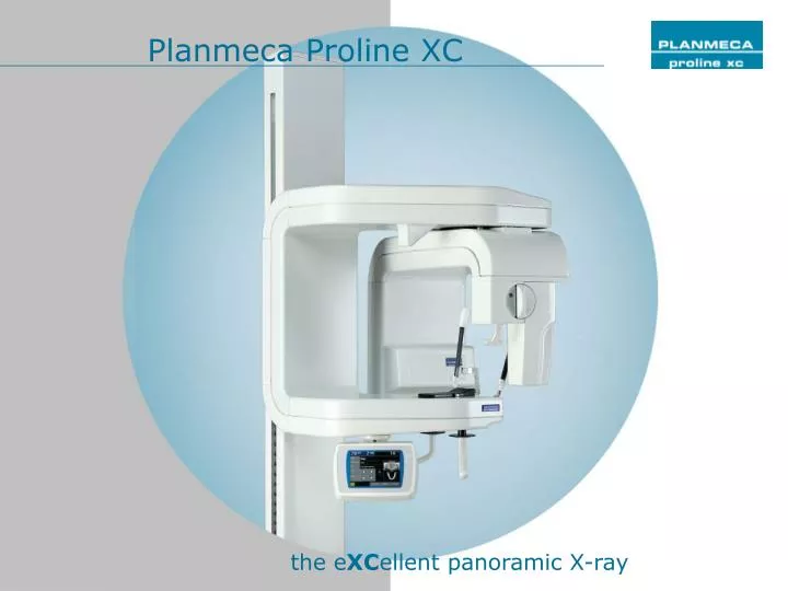
the eXCellent panoramic X-ray Planmeca Proline XC
the best choice for dental office Easy to operate Open and easy patient access Side entry and open view No mirror, no traumatic view of injuries Triple laser beam system for accurate alignment
intuitive controls Graphic User Interface (GUI) for intuitive selection of the wanted program and exposure parameters.
clear access for all patients Wheelchair patients Patient in hospital bed Children can see the accompanying parent
clear and sharp images The focal layer shape is based on scientific study on human jaw shape Planmeca Proline XC images have constant magnification
clear and sharp images Jaw shape and size vary according to age, gender and race Adjustable focal layer to fit different patient’s anatomy size: small – medium - large shape: narrow – average – square
clear and sharp images • The path of the rotating centre of the radiation beam influences the image quality • Long path starting and ending well outside the jaw • less radiation • less shadows • Planmeca Proline XC images have no ghost shadows • high diagnostic quality even in cases with metallic items in the object
clear and sharp images Elimination of the cervical column shadow reduced rotation speed at the central incisor region uniform darkness, and contrast Constant potential (DC) generator less radiation to patient improved contrast reproducible images Constant potential x-ray generation kV t Conventional x-ray generation kV t
successful images every time continuous adjustment measure Film: AEC Automatic Exposure Control • anatomic structures vary between patients • automatic selection of exposure parameters • initial measurement • continuous adjustment • selection of target film darkness (optical density) level Digital: AGC Automatic Gain Control • Optimizes the image quality by adjusting the sensitivity of the sensor according to the amount of incoming radiation. • Prevents pixel saturation even in soft tissue and direct radiation areas
high-tech digital imaging system Planmeca Dimax Combined digital panoramic and cephalometric system Digital sensor employs latest CCD technology Extremely fast image information transfer Eliminates the use of film, film processor and darkroom the most advanced digital panoramic X-ray system available
high-tech digital imaging system Dimax sensor Sensitive area: 9 x 136 mm Pixel size: 33 µm (binning: 66 / 99 / 132 µm) Short decay time scintillator Very high signal to noise ratio (SNR) Very high detective quantum efficiency (DQE) Dynamic range: 17 bit, 131072 grey values Data transfer: fast data link (10 MB/s) between sensor and computer excellent images real time imaging
high-tech digital imaging system Dimaxis Pro software Real-time image acquisition Image enhancement Contrast / darkness Flashlight Multiple filters Measurements Annotations all in one dental imaging Planmeca Dixi®3 Intracam DICOM compatibility meeting all ADA service level requirements DICOM Basic DICOM Advanced Planmeca TWAIN
functional exposure programs Standard panoramic program Paediatric program • reduced area from sides • 20% less patient radiation dose
functional exposure programs Vertical segmenting • limited exposure of the diagnostically interesting area • minimum radiation to patient • reduces the patient dose up to 80%
functional exposure programs Automatic TMJ program TMJ views with mouth closed and open on one film Maxillary sinus program Special focal layer
easy cephalometry Proline CM Cephalostat • automatic alignment of the tube head • motorised aperture selection • soft tissue filter positioning from control panel • Can also be added to the Planmeca Proline XC unit any time in the future
easy cephalometry lateral AP/PA submento-vertex water’s Proline Cephalostat • Best cephalometric system for all radiographic projections towne’s
easy cephalometry Dimax Cephalostat • Same movable digital sensor for panoramics and cephalometry • Optionally two fixed digital sensors • Field height 23 cm • Field width 18 - 27 cm (adjustable) • image field cover also the back of the skull • image size according to diagnostic need • reduced radiation to patient
easy cephalometry Horizontal scanning • the sensor moves horizontally (Scanning time 17 s) • X-ray beam turns around the focus • magnification equal in both directions = no distortion • allows field width of up to 29 cm = whole skull
easy cephalometry • Soft tissue filtration done in the Dimaxis imaging software • no need for a mechanical filter • possible to view the image with and without the filter
secure image storage Solid database • Integrated professional database for image storage and administration • Long-term archiving • Automatic recovery system • Built-in warning system • Automated back-up • secure storage of images • no lost information even if computer system crashes
computer system requirements SERVER PC (recommended) Processor: Pentium III RAM: 256 MB or more Hard Disc: 2 x 80 GB Local bus: PCI, USB CD-ROM drive Backup medium: MO disc, DAT or other Operating system: Windows 2000, XP, 2003 CLIENT PC (minimum) Processor: Pentium III RAM: 256 MB or more Hard Disc: 50 Mb free space Local bus: PCI CD-ROM drive Operating system: Windows 2000, XP, 2003
film marking Autoprint • Automatic film marking during exposure • All necessary information • patient ID • clinic/doctor ID • exposure parameters • date, time and film orientation • no more mixed images • easy to reproduce images at later times • easy and systematic way to document images
film marking Admark • Film marker for any green sensitive film • Used with Autoprint or as stand alone • All necessary information • patient ID • clinic/doctor ID • exposure parameters • date, time, ceph projection and film orientation • no more mixed images • easy to reproduce images at later times • easy and systematic way to document images
Moreinformation: Mr. Erkki Hiltunen Product Manager, X-ray Division tel: +358 20 7795 456 erkki.hiltunen@planmeca.com www.planmeca.com Mrs. Sari Mäkinen Product Specialist, X-ray Division tel: +358 20 7795 469 sari.makinen@planmeca.com the end