Download

1 / 31
480 likes | 2.47k Views
ANATOMY OF PLEURA. Dr. Mujahid Khan. Location. The pleurae and lungs lie on either side of the mediastinum within the chest cavity Each pleura has two parts: Parietal layer Visceral layer. Parietal Layer. It lines the thoracic wall

E N D
ANATOMY OF PLEURA Dr. Mujahid Khan
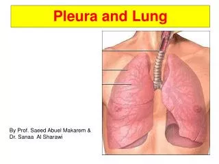
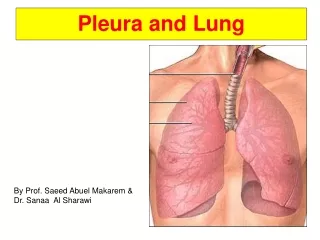
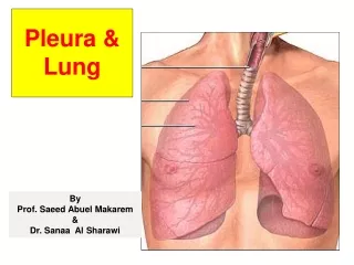
Location • The pleurae and lungs lie on either side of the mediastinum within the chest cavity • Each pleura has two parts: • Parietal layer • Visceral layer
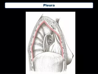
Parietal Layer • It lines the thoracic wall • Covers the thoracic surface of the diaphragm and the lateral aspect of the mediastinum • Extends into the root of the neck to line the undersurface of the suprapleural membrane at the thoracic outlet
Visceral Layer • It completely covers the outer surfaces of the lungs • Extends into the depths of the interlobar fissures
Pleural Cuff • The two layers continuous with one another by means of a cuff of pleura • This cuff surrounds the structures entering and leaving the lung at the hilum of each lung • Pleural cuff hangs down as a loose fold called the pulmonary ligament
Pleural Cavity • The parietal and visceral layers are separated from one another by a slitlike space called pleural cavity • Clinicians use the term pleural space instead of the anatomic term pleural cavity • Pleural cavity contains thin film of tissue fluid called pleural fluid • Fluid permits the two layers to move on each other with the minimum of friction
Cervical Pleura • Parietal pleura is divided into the region in which it lies or the surface that it covers • The cervical pleura extends up into the neck • It lines the undersurface of the suprapleural membrane • It reaches a level 1 to 1.5 in. (2.5 to 4 cm) above the medial third of the clavicle
Costal Pleura • It lines the inner surfaces of: • The ribs • The costal cartilages • The intercostal spaces • The sides of the vertebral bodies • The back of the sternum
Diaphragmatic Pleura • It covers the thoracic surface of the diaphragm • In quiet respiration, the costal and diaphragmatic pleurae are in apposition to each other below the lower border of the lung • Costal and diaphragmatic pleurae separate in deep inspiration
Costodiaphragmatic Recess • The lower area of the pleural cavity into which the lung expands on inspiration is referred to as the costodiaphragmaticrecess
Mediastinal Pleura • It covers and forms the lateral boundary of the mediastinum • It is reflected as a cuff around the vessels and bronchi at the hilum of the lung • Then continuous with the visceral pleura • Each lung lies free except at the hilum • it is attached to the blood vessels and bronchi that constitute the lung root
Mediastinal Pleura • During full inspiration the lungs expand and fill the pleural cavities • During quiet inspiration the lungs do not fully occupy the pleural cavities at four sites • The right and left costodiaphragmatic recesses • The right and left costomediastinal recesses
Costodiaphragmatic recesses • Are slitlike spaces between the costal and diaphragmatic parietal pleurae • Separated only by a capillary layer of pleural fluid • During inspiration, the lower margins of the lungs descend into the recesses • During expiration, the lower margins of the lungs ascend so that the costal and diaphragmatic pleurae come together again
Costomediastinal Recesses • Are situated along the anterior margins of the pleura • They are slitlike spaces between the costal and the mediastinal parietal pleurae • Separated by a capillary layer of pleural fluid • During inspiration and expiration, the anterior borders of the lungs slide in and out of the recesses
Nerve Supply • The parietal pleura is sensitive to pain, temperature, touch and pressure, and is supplied as follows: • The costal pleura is segmentally supplied by the intercostal nerves • The mediastinal pleura is supplied by the phrenic nerve • The diaphragmatic pleura is supplied over the domes by the phrenic nerve and around the periphery by the lower six intercostal nerves
Nerve Supply • The visceral pleura covering the lungs is sensitive to stretch • It is insensitive to common sensations such as pain and touch • It receives an autonomic nerve supply from the pulmonary plexus
Pleural Fluid • The pleural space normally contains 5 to 10 ml of clear fluid • It lubricates the opposing surfaces of the visceral and parietal pleurae during respiration • The formation of the fluid results from hydrostatic and osmotic pressures between the capillaries • The pleural fluid is normally absorbed into the capillaries of the visceral pleura
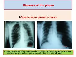
Pleural Fluid • Any condition that increases the production of the fluid or impairs the drainage of the fluid results in the abnormal accumulation of fluid, called pleural effusion • The presence of 300 ml of fluid in the costodiaphragmatic recess in an adult is sufficient to enable its clinical detection • The clinical signs include decreased lung expansion on the side of the effusion, with decreased breath sounds and dullness on percussion over the effusion
Pleuricy • Inflammation of the pleura secondary to inflammation of the lung called pneumonia • Pleural surfaces become coated with inflammatory exudate, causing the surfaces to be roughened • Produces friction, and a pleural rub • It can be heard with the stethoscope on inspiration and expiration
Pleuricy • Often the exudate becomes invaded by fibroblasts • That lay down collagen and bind the visceral pleura to the parietal pleura • Forms pleural adhesions