Download
1 / 42
420 likes | 436 Views
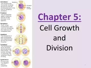
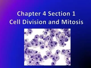
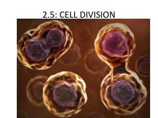
Chapter 10 – Cell Life Cycle. Figure 12.0 Mitosis. Figure 12.1c The functions of cell division: Tissue renewal. Figure 12.2 Eukaryotic chomosomes. Figure 12.3 Chromosome duplication and distribution during mitosis.

E N D
Figure 12.3 Chromosome duplication and distribution during mitosis
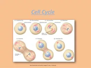
Figure 12.5 The stages of mitotic cell division in an animal cell: G2 phase; prophase; prometaphase
Figure 12.5 The stages of mitotic cell division in an animal cell: metaphase; anaphase; telophase and cytokinesis.
Figure 12.7 Testing a hypothesis for chromosome migration during anaphase
Figure 12.10 Bacterial cell division (binary fission) (Layer 3)
Figure 12.17 The growth and metastasis of a malignant breast tumor
Figure 12-17x2 Mammogram: normal (left) and cancerous (right)
Figure 13.x2 Human female chromosomes shown by bright field G-banding
Figure 13.x3 Human female karyotype shown by bright field G-banding of chromosomes
Figure 13.x4 Human male chromosomes shown by bright field G-banding
Figure 13.x5 Human male karyotype shown by bright field G-banding of chromosomes
Figure 13.5 Three sexual life cycles differing in the timing of meiosis and fertilization (syngamy)
Figure 13.6 Overview of meiosis: how meiosis reduces chromosome number
Figure 13.9 The results of alternative arrangements of two homologous chromosome pairs on the metaphase plate in meiosis I