Download
1 / 28
300 likes | 378 Views
Learn about antibody-mediated cell dysfunction and complement-mediated inflammation in Type II Hypersensitivity, as well as IgG or IgM involvement in complement-mediated tissue destruction in Type III Hypersensitivity. Examples include AIHA, ABO incompatibility, and autoimmune diseases like Graves' disease. Discover testing methods and mechanisms of cellular destruction.

E N D
Immunology/ Pharmacy Students • Hypersensitivity (2) Dr. Mohammad Odibate Department of Microbiology and immunology Faculty of Medicine, Mu’tah University
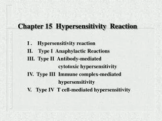
Hypersensitivity reactions= Policeman and the thief Ag Type 2 hypersensitivity Type 3 hypersensitivity
Type II Hypersensitivity • General characteristics: • Involved antibodies: IgG and IgM • Triggering antigens • Foreign antigens • Drug or bacterial induced altered self antigens Soluble Ab Complement mediated tissue destruction Cell with Ag on its surface
Type II Hypersensitivity Type II Hypersensitivity is subdivided into three ways Ab mediated cell dysfunction (no cell depletion, destruction, or inflammation) Target cell depletion or destruction without inflammation Complement mediated Inflammation
Type II Hypersensitivity 1. Target cell depletion or destruction without inflammation • Examples • Autoimmune Hemolytic Anemia (AIHA) • Autoimmune Neutropenia • Erythroblastosisfetalis • ABO incompatibility • Mechanism of cell destruction is mediated by • Antibodies • Complement • Natural killer (NK) cells
Erythroblastosis Fetalis Rh- mother Placenta Anti Rh Antibodies (IgG type) Second Rh+ fetus First Rh+ fetus Rh+ antigens First pregnancy Between pregnancies Second pregnancies
ABO incompatibility • Are examples of cellular destruction that result from antibody combining with blood antigens • Acute hemolytic transfusion reactions may occur within minutes or hours after receipt of incompatible blood Donor RBCs destruction = +
Type II Hypersensitivity 1. Target cell depletion or destruction without inflammation • Autoimmune Hemolytic Anemia (AIHA) • Causes of AIHA: • Idiopathic • Drug or bacterial induced altered self antigens • RBCs with altered self antigens Bacteria RBC RBCs will be destructed by either complement activation or ADCC Bacterial Ag Self Ag Cell RBC ADCC Drug
Antibody dependent cell mediated cytotoxicity (ADCC) APC Present RBC proteins to T cells RBC • RBCs with altered self antigens APC uptakes cellular debris from destructed RBCs Antibodies will be produced ,released into circulation, and Eventually bind to the intact RBCs in circulation holding the same altered antigen cell NK cell Perforin and granzymes NK has a receptor for the antibody Fc portion RBC RBC
Type II Hypersensitivity 2. Complement mediated Inflammation Mechanism Ab directed against kidney basement membrane = complement mediated destruction. Complement proteins mediate neutrophilechemotaxis to the site of Ab activation. 1. Goodpasture's syndrome Examples Kidney with Goodpasture's syndrome Normal Kidney Filtration Basement membrane (BM) Ab + complement= BM destruction Renal damage (hematuria) Urine
2. Acute Rheumatic Fever M protein S. pyogenes M-protein mimics the self proteins present in heart, CNS, and joints • Anti-M antibodies start fighting • against bacteria • Anti-M antibodies also cross react with • self Ag and mediate Immune destruction Protection Autoimmunity Autoimmunity
Type II Hypersensitivity 3. Ab mediated cell dysfunction (no cell depletion, destruction, or inflammation) Examples Graves' disease TSH Abnormal Normal Thyroxin he disorder results from an antibody, called thyroid stimulating immunoglobulin (TSI), that has a similar effect to thyroid stimulating hormone (TSH). These antibodies cause the thyroid gland to produce excess thyroid hormone
Type II Hypersensitivity 2. Myasthenia gravis Autoantibody produced against acetylcholine (Ach) receptors which inhibits the Ach mediated muscle contraction leading to muscle weakness
Type II Hypersensitivity Testing for type AIHA Direct Coombs’ antiglobulin testing (DAT) The direct antiglobulin test (DAT) detects sensitized red cells with IgG and/or complement components in vivo. • In vivo coating of red cells • Example: • Patient with suspected AIHA • Rh hemolytic disease of the newborn
Type II Hypersensitivity Testing for type II Hypersensitivity 2. The indirect Coombs’ antiglobulin testing: Detects anti-erythrocytes Ab in serum in vitro Patients serum Anti-human (Coomb’s reagent) Patient’s blood is positive for anti-RBC Abs
Type III Hypersensitivity • General Characteristics • Antibodies involved • IgG or IgM is involved and destruction is complement mediated • The antigens are: • Soluble • Endogenous (SLE) or exogenous (Bacterial, viral, or fungal) • Not intrinsic to tissue • Sites in which this typically occurs include the glomerular basement membrane vascular endothelium, joint linings, and pulmonary alveolar membranes Type III Soluble Ag Soluble Ab Complement mediated tissue destruction Tissue
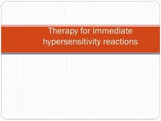
Type III Hypersensitivity Types 1. Generalized reaction (Serum Sickness reaction) Antibodies and medications are useful under normal conditions: Anti-tatanus Anti-rabies Treating Clostridium tetaniand Rabies infections Snake antisera Neutralizing snake venom Certain medications Treating certain diseases But these might have negative effect through the induction of type III Hypersensitivity Anti-tatanus Anti-rabies Type III Hypersensitivity Snake antisera Certain medications
Type III Hypersensitivity Types 1. Generalized reaction (Serum Sickness reaction) Anti-tatanus Soluble protein Anti-Anti tetanus Plasma cell Immune response against anti-tetanus Insoluble Ab-Ab complex Recruitment of inflammatory cells Destructive enzymes Complement activation
Type III Hypersensitivity Types Generalized reaction (Serum Sickness like reaction). • Fever • Generalized Lymphadenopathy • Rashes • Proteinuria and hematuria • Polyarthritis • Pericardial pain • Multiple vasculticleasions Recruitment of inflammatory cells Destructive enzymes Complement activation
Type III Hypersensitivity Types • Serum sickness like reaction : • Rhumatoid arthritis: is an IgM produced against own IgG which deposit in joints. The IgM is called Rhumatoid factor • Polyarthritisnodosa: antibody produced against HBsAg • Post streptococcal glumerulonephritis: after streptococcal infection the Ags of bacteria are circulated in blood then the body will start producing Abs that both love to deposit in kidney • Reactive arthritis: After GIT infection by Yersinia,the bacterial Ags will deposit in the joints with their corresponding Ab which cause inflammation in joints
Type III Hypersensitivity Types 2. Autoimmune Diseases • Systemic lupus erythematosus (SLE) • Type III hypersensitivity reactions can be triggered by antibodies directed against self antigens such as DNA and nucleohistones • Immune complex deposition involves multiple organs, but the main damage occurs to the glomerular basement membrane in the kidney • B. Rheumatoid arthritis • An antibody called rheumatoid factor is directed against IgG. • Immune complex deposition occurs in the membranes of inflamed joints. Complement enhances tissue destruction in both diseases.
Systemic lupus Erythematosus (SLE) SLE as a type II hypersensitivity SLE as a type III hypersensitivity Autoantibody produced against intrinsic self antigens Autoantibody reacted with soluble self antigens originated from damaged cells Endogenous antigens Autoantibody + self antigen (intrinsic) Cell Autoantibody Immune complex Cell destruction Precipitation in tissues Complement fixation and tissue damage soluble self antigens originated from damaged cells
Type IV Hypersensitive reaction • DTH response is from: • Th1 cells release cytokines to activate macrophages causing inflammation and tissue damage. • Continued macrophage activation can cause chronic inflammation resulting in tissue lesions, scarring, and granuloma formation.
Type IV Hypersensitive reaction Stages of Type IV DTH 1- Sensitization stage • Th1 cells against DTH antigens are generated by dendritic cells during the sensitization stage. • These Th1 cells can activate macrophages and trigger inflammatory response.
Type IV Hypersensitive reaction 2- Effector stage Stages of Type IV DTH • Th1 memory cells are activated and produce cytokines. • IFN-g, TNF-a, and TNF-b which cause tissue destruction, inflammation. • IL-2 that activates T cells and CTLs. • Chemokines- for macrophage recruitment. • IL-3, GM-CSF for increased monocyte/macrophage • Inflamed area becomes red and fluid filled can form lesion. • Continued exposure to antigen can cause chronic inflammation and result in granuloma formation.
Type IV Hypersensitive reaction Stages of Type IV DTH
How to remember the types of hypersensitivity? A B C D I= Allergic Anaphylaxis Atopy II= antiBody III= immune Complex IV= Delayed A C I D naphylaxis = Type I ytotoxic = Type II Acid mmune complex = Type III elayed = Type IV