Download
1 / 28
310 likes | 514 Views
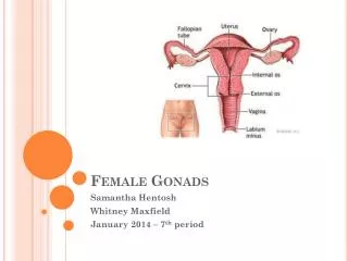
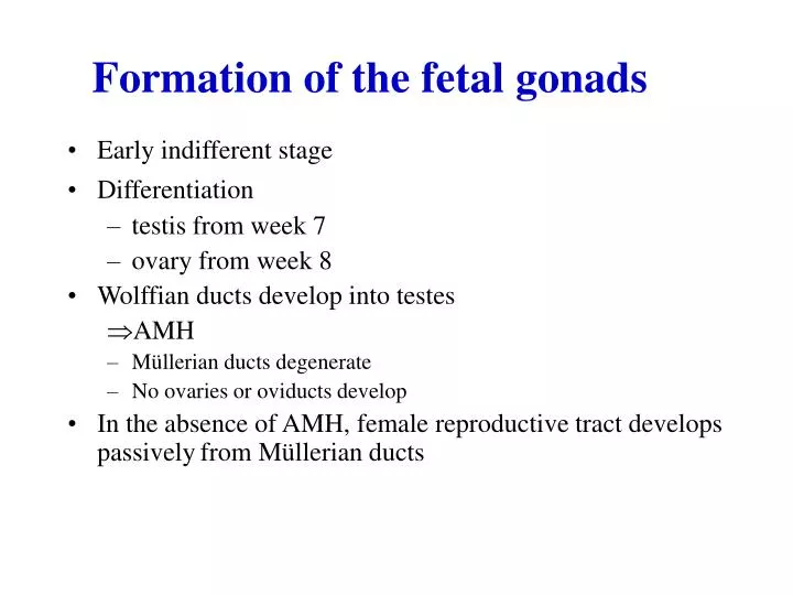
Formation of the fetal gonads. Early indifferent stage Differentiation testis from week 7 ovary from week 8 Wolffian ducts develop into testes AMH Müllerian ducts degenerate No ovaries or oviducts develop

E N D
Formation of the fetal gonads • Early indifferent stage • Differentiation • testis from week 7 • ovary from week 8 • Wolffian ducts develop into testes • AMH • Müllerian ducts degenerate • No ovaries or oviducts develop • In the absence of AMH, female reproductive tract develops passivelyfrom Müllerian ducts
I. Development of the human male reproductive tract The male accessory genital organs are complex. Three different structures participate and each of them undergoes essential transformations: 1. The wolffian ducts constitute the epididymis, the seminal vesicles, the ejaculatory ducts and the central part of the prostate. 2. The müllerian ducts regress in the male and form occasionally the testicular appendices. 3. The urogenital sinus forms the peripheral and transitional zones of the prostate
Male Reproductive System The male reproductive system is composed of 3 main components: the testes, the duct system and the accessory glands.
I.The testis In the adult male, the average testis is approximately 1.5 to 2 inches long. II. Seminiferous tubules
III. The ex-current ducts of the testis The contents of the rete testis, namely, spermatozoa and testicular fluid, pass directly through a series of efferent ductules (ductuli efferentes), into the main ex current duct, the epididymis. I. Structure of the adult epididymis The epididymis lies adjacent to the testis, and is a crescent shaped organ. The proximal end of the epididymis leads to the ductuli efferentia (vasa efferentes), or efferent ducts, along which spermatozoa pass from the rete canal of the testis. The distal end of the epididymis is continuous with the vasa deferentia which eject spermatozoa from the body at ejaculation.
Vas Deferens The vas deferens are a pair of ducts with one leading from the distal end of the cauda of each epididymis. Spermatic cord The spermatic cord connect the testis to its life support mechanisms, the convoluted testicular arteries and surrounding venus plexus, and nerve trunks Ejaculatory duct Two ejaculatory ducts are short tubes that pass through the prostate gland to terminate in the urethra Seminal vesicles The seminal vesicles are lateral expansions of the vas deferens. They derive from the wolffian ducts, and their aspect is that of an irregular gland of 5-8 cm
Accessory Glands The accessory glands are located along the pelvic portion of the urethra, with ducts which empty their secretions into the urethra. They include the vesicular glands, the prostate gland and the bulbourethral glands
Function of the testis I. Secretion of Testosterone II. Spermatogenesis
Spermatogenesis can be divided into three parts: • Spermatocytogenesis • proliferative phase. • B).Meiosis—production of the • haploid gamete • C) Spermiogenesis • Spermatids mature into • spermatozoa (sperm)
Events associated with spermiogenesis: a. Condensation of nuclear material b. Removal of extraneous cytoplasm c. Formation of the acrosome d. Formation of tail structures 3. Spermiogenesis ends in the testis with release of the spermatozoa from the Sertoli cell
Different parts of the Spermatozoa: • Head • Neck • Middle piece • Principal piece • End piece
Hormonal control of spermatogenesis(male reproductive tract) • 1. Hormones that affect spermatogenesis • Testosterone • i. Produced by Leydig cells • ii. Promotes Sertoli cell function • . • Estradiol • i.Produced by Sertoli cells and it is essential for spermiation • Inhibin • i. Produced by Sertoli cells
d. FSH—follicle stimulating hormone • i. Produced by anterior pituitary • FSH regulates the mitotic divisions and efficiency of type A spermatogonia development • FSH controls entry of stem cell type A spermatogonia into proliferating pool • iv) The yield of spermatozoa is increased by FSH • V) FSH is necessary during development
. • LH—lutenizing hormone • i. Produced by anterior pituitary • ii. Increases testesterone production by Leydig cells • GnRH—gonadotropic releasing hormone • Hypothalamic hormone that controls release of anterior pituitary hormones (LH and FSH) • Once the Sertoli function is developed, testosterone alone will maintain spermatogenesis • The yield of spermatozoa is increased if FSH is present
Non-hormonal factors a) Temperature The optimal temperature for spermatogenesis is 34-35 C. b) Diet . Diet must contain vitamin A, B12, folic acid and vitamin C and E which is essential for spermatogenesis.
Function of Sertoli cells 1) It prevent the antibody producing cells in Extra cellular fluid from reaching the tubular sperm factory, thus preventing the formation of antibodies against the highly differentiated spermatozoa. 2) It provides a mechanical support for seminiferous tubules. 3) It provides nutrition for the developing sperm 4) Sertoli cells have an important phagocytic function. They engulf the cytoplasm extruded from the spermatids during their remodeling and destroy defective germ cells that fail to successfully complete all stages of spermatogenesis.
5) Sertoli cells secret fluid into the seminferous tubule whichpushes and flushes the release sperm from tubule into the epididymis for storage. 6) Sertoli cells secrete an androgen binding protein (ABP) that binds testosterone thus maintaining a very high level of this hormone which is essential spermatogenesis. 7) Sertoli cells secret inhibin to regulate FSH secretion. Also Mullerian inhibitory factor during fetal life to inhibit the formation of Fallopian tube from Mullerian duct.
Function of Testosterone A) Prenatal: during fetal development B) At puberty: 1) Development of male sex organs: External genitalia: Penis increases in width and length 8 fold until age of 20. Enlarged testes and scrotum. Internal genitalia: Enlarged seminal vesicle and begin to secret and form fructose, also enlarge of prostate and bulbourethral glands.
1) Development of secondary sex characters Beginning at puberty and ending at maturity Hair growth and distribution of body hair: Voice: Testosterone causes enlargement of larynx, and voice become deeper. Body conformation: Shoulders broaden, narrow pelvis and muscles enlarge. Skin: Sebaceous gland secretion increases leading to acne. 1) Anabolic effect of Testosterone Testosterone increase the synthesis and decreased the breakdown of proteins leading to increase in growth.
4) Effect on bone growth and calcium retention It causes growth of long bones (increase to about 3 inches per year). 5)Effect on the red blood cells The average man has about 700,000 more RBCs per mm3 than the average women and this is due to increased metabolic rate of bone marrow 6) Effects on basal metabolism Testosterone increase the basal metabolic rate (BMR) as much as 5-10% above normal, which is due to protein anabolism.
SEMEN: Sperm pass through the vas deferens and connect to a short ejaculatory duct that connects to the urethra. The urethra passes through the penis and opens to the outside. Secretions from the seminal vesicles (70%) add fructose and prostaglandins to sperm as they pass. The prostate gland (20%) secretes a milky alkaline fluid. 7% of spermatozoa, epididymal fluids and the bulbourethral gland (3%) secretes a mucus-like fluid that provides lubrication for intercourse. Sperm and secretions make up semen. Average PH of the semen is about 7.5, color is milky and apperance is mucoid
Sperms Terms: Normospermia: Oligospermia: Azoospermia: Aspermia:
Male sex act consists of 3 parts: • 1) Erection:It is caused by Parasympathetic impulses that pass from the sacral portion of the spinal cord to the penis. • 2) Emission: • Emission begins with contraction of the muscular coat of the prostate. After that vas deferens to cause expulsion of sperm into the internal urethra, then followed by contraction of the seminal vesicles fluid.
3)Ejaculation. 1) Contraction of the ischiocavernosus and bulbocavernosus muscles that compress the bases of the penile erectile tissue. 2) wavelike increases in pressure in the genital duct and urethra which ejaculate the semen from the urethra to the exterior. This entire period of emission and ejaculation is known as male orgasm. At the end of these process the male sexual excitement disappears almost within 1-2 minutes, and erection ceases.