Download
1 / 22
220 likes | 407 Views
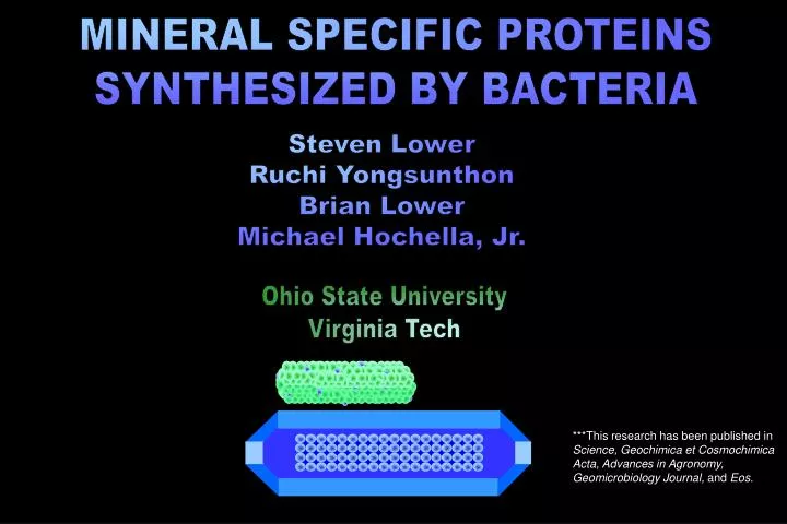
MINERAL SPECIFIC PROTEINS SYNTHESIZED BY BACTERIA. Steven Lower Ruchi Yongsunthon Brian Lower Michael Hochella, Jr. Ohio State University Virginia Tech.

E N D
MINERAL SPECIFIC PROTEINS SYNTHESIZED BY BACTERIA Steven Lower Ruchi Yongsunthon Brian Lower Michael Hochella, Jr. Ohio State University Virginia Tech ***This research has been published in Science, Geochimica et Cosmochimica Acta, Advances in Agronomy, Geomicrobiology Journal, and Eos.
400 nm bacterium-mineral interface interfacial forces and proteins *forces control, and are themselves modulated by, the expression of biopolymers on a bacterium’s surface G. Bowles
Microorganisms Humans • # of cells • # of species • # years • 1030 109 • 106 100 • 109 106 Consider the following… A comparison of two organisms that work in the realm of the nanometer.
Shewanella interactions with Fe oxyhydroxides metal reducing bacteria & oxides • primitive form of respiration using Fe(III) solids • remediation of organic and inorganic pollutants in surface and subsurface environments Shewanella Fe(III) mineral
Fe(III) oxide H+ H+ e- O2 H2O NADH NAD+ ADP ATP outside outer membrane periplasm inner membrane inside
pH Mr force microscopy 2D gel electrophoresis cell lithography phage display library Cell-material interface discover forces and proteins F~e -D theory mimic and utilize information
living cell biological force microscopy laser lever mineral
G x m1 x m2 (distance)2 Force-distance curves using different minerals, bacteria, and solutions approach retraction
H R 6 d 2 electrostatic and van der Waals forces DLVO Theory 4 p s1 s2 R e - kd F(d) = e eo k s = surface charge R = radius e = dielectric constant eo= vacuum permittivity d = distance k = 1 / Debye length H= Hamaker constant R = radius d = distance ~(salt concentration) –1/2
subsurface transport in the environment (approach measurements between G- bacterium and a silicate) green – low IS blue – high IS approach only
Force-distance curves (retraction forces between Shewanella and AlOOH vs FeOOH) approach retraction Shew AlOOH FeOOH
diaspore goethite (AlOOH) (FeOOH) + 39 7 - 26 6 aerobic + + - - anaerobic + 41 4 - 137 20 energy values between Shewanella and mineral (as function of oxygen concentration) attoJoules (10-18 J) Control experiment with nonviable cells ~6 aJ (did not change with mineral, oxygen concentration, or contact time)
Fe(III) oxide outside outer membrane H+ periplasm e- H+ inner membrane O2 H2O NADH NAD+ inside ADP ATP
d = distance or extension k = Boltzmann’s constant T = temperature b = persistence length (0.38nm Ca-Ca in protein) L = contour length (length of stretched protein chain) Protein folding/unfolding Worm-like Chain Model F(d) = (k T / b) [0.25 (1 – d / L)–2 – 0.25 + d / L]
OM protein expression patternsShewanella andAlOOHorFeOOH protein signature observed in 80% of the data; only with goethite after some period of “recognition time”
2D Gel Electrophoresis of OM Proteins pH Fe+3 O2 Mr pH Mr excise proteins & fragment into peptides mass spec & database search extract membrane proteins compare with force signature culture cells separate proteins force microscopy
terminal electron acceptor -O2vsFe(III) kDa 150 100 75 50 37 25 pH 4.0 5.0 6.0 7.0 8.0 9.0
Cell-material interface pH Mr force microscopy 2D gel electrophoresis cell lithography phage display library discover forces and proteins F~e -D theory mimic and utilize information
peptide phage library expose to target mineral wash away unbound phage elute bound phage “bio-panning” isolate clones, sequence DNA to find mineral-binding motif,
protein cell substrate biological cell lithography biological cell lithography bacterium as living “pen” that produces and secretes genetically engineered proteins S-layer protein T. Beveridge
bacterium-mineral interface nanoscale forces and proteins • quantify natural forces of affinity between inorganic crystalline phases and proteins synthesized by bacteria • use theoretical models and protein expression patterns to identify putative mineral specific proteins • mimic natural specificity by attempting to fabricate peptides with unique mineral-binding motifs • use living microbial cells as a lithographic tool
Acknowledgements Acknowledgements Terry Beveridge, John Smit, Courtney Crummett, Graeme Bowles National Science Foundation (GEO & ENG) Department of Energy American Chemical Society ***This research has been published in Science, Geochimica et Cosmochimica Acta, Advances in Agronomy, Geomicrobiology Journal, and Eos. Steven Lower – Ohio State University – Lower.9@osu.edu