Download
1 / 48
1k likes | 4.44k Views
Cestodes. Gregory L. Brower, D.V.M., Ph.D. Cell Biology and Anatomy School of Medicine Columbia, South Carolina. Cestodes (Tape Worms). Taenia solium (pork tapeworm) - Cysticercus Taenia saginata (beef tapeworm) Diphyllobothrium latum (fish tapeworm)

E N D
Cestodes Gregory L. Brower, D.V.M., Ph.D. Cell Biology and Anatomy School of Medicine Columbia, South Carolina
Cestodes (Tape Worms) • Taeniasolium(pork tapeworm) - Cysticercus • Taeniasaginata(beef tapeworm) • Diphyllobothriumlatum (fish tapeworm) • Echinococcusgranulosus (unilocularhydatid) • Echinococcusmultilocularis (alveolar hydatid) • Hymenolepis nana (dwarf tapeworm) • Hymenolepisdiminutia • Dipylidiumcaninum
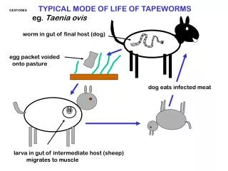
General Structure of Tapeworm • Head region (scolex) : contains suckers and hooks used to attach to a host organism. • Proglottids : square body segments used for reproduction.
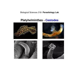
Tapeworm Structure • Scolex - Attachment organ • Zone of Proliferation - Undifferentiated area behind the scolex (neck region) • Strobilia - Chain of segments (proglottids) • Immature proglottids - developing reproductive • Mature proglottids: mature reproductive organs. • Gravid proglottids: contain eggs in the uterus.
Immature Segment note that the reproductive organs are just beginning to differentiate. • (Carmine stained) Developing reproductive organs
Mature Segments (Proglottids) Tapeworms are Hermaphroditic
Taeniasis:Geographic Distribution • Worldwide, depending on dietary habits, and quality of cattle and pork farming.
Taenia solium (Pork Tapeworm)Morphology Adult Worm: 2-4 m long, 700-1000 segments: Scolex Neck Immature segment Mature segment Gravid segment
Cysticercus cellulosae It is soybean-like in shape, with the small scolex invaginated into the translucent cyst (left). The scolex evaginated from the cyst (right). Cysticercius cellulosae
Taenia eggs The eggs of Taenia saginata and T. Solium are morphologically indistinguishable.
Abdominal discomfort, epigastric pain, vomiting Physical presence of the worm Gastro-intestinal Site Symptoms Pathogenesis Other organs (cysticercosis) Cyst formation in brain, eye, lung and liver may cause related symptoms Physical mass and inflammation Taeniasis:Symptoms
Taeniasis:Diagnosis Symptoms History of eating undercooked beef or pork Recovery of proglottids and/or eggs in the stool
Cysticercosis:Diagnosis • CNS and/or symptoms involving other organs • History of ingesting food with T.soliumeggs • Radiographic localization of cysticercal lesions in tissues
Cysticercosis:Disease Cerebral cysticercosis
Cysticercosis:Disease Cardiac cysticercosis Ocular cysticercosis
Taeniasis:Treatment and Prevention Treatment • Praziquantel; scolex expulsion is essential Prevention • Adequate cooking of meat • Freezing meat below 10º C
Fish TapewormDiphyllobothriumlatum Distributed worldwide: freshwater (great lakes)
1 cm Diphyllobothrium latum: Morphology
Diphyllobothriasis:Symptoms Site Symptoms Pathogenesis gastro-intestinal abdominal discomfort, rarely severe cramping pain, diarrhea alternated by constipation, vomiting related to worm burden weight loss, vitamin B-12 deficiency physical mass and inflammation general
Diphyllobothriasis:Diagnosis • symptoms • history of eating raw fish • recovery of proglottids and/or eggs in the stool
Diphyllobothriasis:Treatment • Praziquantel is the drug of choice • Avoid uncooked fish from infested waters • Freezing for 24 hours and pickling kills the tape
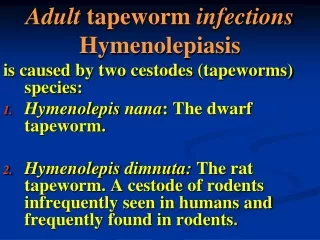
Hymenolepis nana (Dwarf Worm)Symptoms • Lighter infections: mild abdominal disturbance • Heavier infections: enteritis
Hymenolepis nana (Dwarf Worm)Diagnosis, Treatment And Control Diagnosis rodent infestation ova in the feces Treatment and Control Praziquantel is the drug of choice rodent population control
Echinococcusgranulosus(hydatid)Case History • 32-year-old Kenyan woman presented in Boston with a 3-month history of cough that was initially productive of thick, gray sputum that had gradually become blood-tinged. She had low-grade fevers. Chest radiography revealed an apparent elevation of the right hemidiaphragm. CT scan the chest with contrast medium revealed a low-density cystic mass measuring 13 by 13 by 10 cm in the right lower hemithora.
Echinococcusgranulosus(hydatid)Case History • Ultrasound-guided aspiration of the contents of the cyst yielded clear, colorless fluid containing a protoscolex of Echinococcusgranulosus. Three days later, cystectomy was performed, with the use of specific precautions to prevent local spread of disease. After adequate antiparasitic treatment, follow-up radiography demonstrated reexpansion of the right lung. The patient has returned to her normal level of activity, and there is no evidence of recurrent disease.
Echinococcus granulosus:Life Cycle http://workforce.cup.edu/buckelew/
abdominal distension, ascitis progressively growing cyst abdomen Site Symptoms Pathogenesis growing cyst liver obstructive jaundice pulmonary abscess, cough, chest pain growing cyst lung Jacksonian epilepsy growing cyst CNS Type-I hypersensitivity Fever, pruritus, urticaria, anaphylactoid reactions Echinococcus granulosus:Symptoms
Echinococcus granulosus:Diagnosis • Endemicity • Symptoms • X-ray and CT scan • Serology • Skin (Casoni) test
Echinococcusgranulosus: Treatment and Control • Surgical removal of the cyst • Praziquantel • Avoidance or treatment of infected canine
Echinococcus multilocularis • Similar to E. granulosus, except: • Secondary hosts and reservoir are rodents. • The egg produces multilocular cysts. • The cestode is more resistant to chemotherapy.
Echinococcusmultilocularis: Treatment and Control • Surgical removal of the cyst • Resistant to praziquantel; Albendazole has some effect • Avoidance control of rodent population