Download
1 / 30
300 likes | 339 Views
Brain Meninges, Ventricles and CSF. Lecture Objectives. Describe the arrangement of the meninges and their relationship to brain and spinal cord. Explain the occurrence of epidural, subdural and subarachnoid spaces.

E N D
LectureObjectives • Describethearrangementofthemeningesandtheirrelationshipto brain and spinalcord. • Explaintheoccurrenceofepidural,subduralandsubarachnoid spaces. • Locate theprincipal subarachnoid cisterns, and arachnoid granulations. • Describetheventriclesofbrainandimportanceoftheirchoroids plexus. • Summarize the pathway of cerebrospinal fluid(CSF) circulation. • Locatethesafesitesforthelumbarpuncture. • Identify brain ventricles in CT scan, MRI andventriculograms.
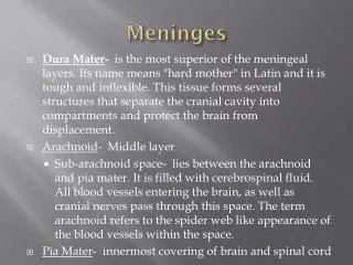
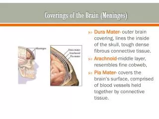
CerebralMeninges • Duramater • Endosteallayer • Meningeallayer • Arachnoidmater • Piamater
DuraMater • Endosteal layer =periosteum • Venoussinuses • Meningeallayer • Continuous with spinaldura mater • Falxcerebri • Shape, Attachments &Sinuses • Tentoriumcerebelli • Shape, Attachments &Sinuses • Tentorialnotch • Falxcerebelli • Shape, Attachments &Sinuses • Diaphragmasellae • Shape, Attachments &Sinuses
DuraMater • Nervesupply • Cranial nerves V &X • Referralpaintotheheadfromabove the tentoriumcerebelli • Spinal nervesC1-C3 • Referralpaintothebackofthehead and neck frombellow tentorium • Sympathetic • Bloodsupply • Internal carotid, maxillary,ascending pharyngeal, occipital & vertebralaa • Middle meningeala.
Dural VenousSinuses • Location • Drains….. • Fate • Superior sagittalsinus • Venouslacunae • Inferior sagittalsinus • Straightsinus • Occipitalsinus • Transversesinus • Sigmoidsinus • Cavernoussinuses • Superior and inferior petrosalsinuses
ArachnoidMater • Subduralspace • Subarachnoidspace • Cerebral BV & cranialnn. • CSF • Subarachnoidcisternae • Arachnoidvilli • Arachnoidgranulations • Fuse with epineuriumat foramina • Except for optic nerve - fusewith sclera
PiaMater • Adheres closely to thebrain • Goes deep into thesulci • Fuse with nervesepineurium • Covers cerebral arteries entering brainsubstance • Specialized over theroofs of ventricles (tela choroidea) • Contributetoformationofchoroidplexuses
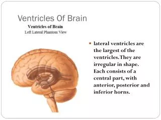
VentricularSystem • Lateralventricles • Interventricularforamina • Thirdventricle • Cerebralaqueduct • Fourthventricle • Centralcanal • Terminalventricle
LateralVentricles • Location • Shape • Parts • Body • Horns • Anterior, posterior,inferior • Choroidplexus
LateralVentricles • Relations • Corpuscallosum • Septumpellucidum • Fornix • Thalamus • Caudatenucleus
ThirdVentricles • Shape • Interventricular foramen(foramina ofMonro) • Cerebral aqueduct (aqueductof Sylvius) • Walls • Anteriorwall • Posteriorwall • Lateralwall • Roof • Floor • Choroidplexus
FourthVentricle • Shape • Relations • Walls • Lateralwalls • Roof (posteriorwall) • Superior and inferior medullaryvelum • Median aperture (foramenof Magendie) • Lateral apertures(foramina of Luschka) • Choroidplexus • Floor (rhomboidfossa)
SubarachnoidCisterns • Extended area ofthe subarachnoidspace • Subarachnoidcisterns • Cerebellomedullarycistern (cisternamagnum) • Medianaperture • Pontinecistern • Lateralapertures • Interpeduncularcistern
Cerebrospinal Fluid(CSF) • 80-150 ml(3-5oz) • Clear liquid containing glucose, proteins, &ions • Functions • mechanicalprotection • floats brain & softens impact with bonywalls • chemicalprotection • optimal ionic concentrations for actionpotentials • circulation • nutrientsandwasteproductstoandfrombloodstream
Origin ofCSF • Choroid plexus =capillariescovered by ependymalcells • 2lateralventricles,onewithineachcerebralhemisphere • Roof of 3rdventricle • Roof of fourthventricle
Drainage of CSF fromVentricles • One median aperture & two lateral apertures allowCSF to exitfromtheinteriorofthebrain
Reabsorption ofCSF • Reabsorbed through arachnoidvilli • grapelikeclustersofarachnoidpenetrateduralvenoussinus • 0.5ml/minreabsorptionrate=sameasproductionrate
Reabsorption ofCSF • Whenpressurein CSF > venous Reabsorptionoccur • When pressure in venous >CSF arachnoid villi work asvalve
Hydrocephalus • Blockage of drainage of CSF (tumor,inflammation, developmental malformation, meningitis, hemorrhage orinjury) • Continuedproductioncauseanincreasein pressure ---hydrocephalus • In newborn or fetus, the fontanels allow this internalpressuretocauseexpansionofthe skull anddamagetothebraintissue • Neurosurgeon implants a drain shunting theCSF to theveinsoftheneckortheabdomen
LeptomeningealDisease • Cancer metastasis throughCSF • Originatefrom • Primary CNStumors • Secondary distant tumors throughblood • Symptomsmayincludeheadache,spineorradicularlimbpainor sensory abnormalities, nausea andvomiting
SubarachnoidHemorrhage • Nontraumatic(spontaneous) • Blood inCSF • Mainly fromaneurisms • Arise at arterial branchpoints • 85% anteriorcirculation • 15% posteriorcirculation • 30% anterior comm., 25% posterior comm.,20% MCA • Themainsymptomisasevereheadachethatstartssuddenly(oftencalled thunderclapheadache) • Traumatic • Morecommon • Duetocontusionsandothertraumaticinjuries • Severeheadache
Blood BrainBarrier • protects cells from sometoxins andpathogens • proteins & antibiotics can notpass but alcohol & anestheticsdo • Structure • tight junctions sealtogether epithelialcells • continuous basementmembrane • astrocyte processescovering capillaries
Blood BrainBarrier • Areas withoutBBB • Area postrema in thefloor of the fourthventricle • Areas in thehypothalamus • Structure • Endothelialfenestrations
Blood Cerebrospinal FluidBarrier • Structure • Endothelialcells • BM of endothelialcells • Palecells • BM of choroidal epithelialcells • Tight junctions seal thechoroidal epithelialcells