Download

1 / 16
160 likes | 231 Views
Explore the layers of the scalp such as skin, connective tissue, and aponeurosis, along with details on meninges, dura mater reflections, and vascular supply to brain structures. Learn about dural venous sinuses, arachnoid layers, and cerebrospinal fluid flow mechanisms.

E N D
Layers of the Scalp • Skin • Connective Tissue • Aponeurosis: Frontalis. Occipitalis. • Loose Areolar Tissue • Pericranium
Cutaneous Nerve Supply • CN V: (Trigeminal Nerve) Ophthalmic, maxillary, mandibular. • Cervical spinal nerves: Ventral rami of C2, C3: Great auricular nerve (C2,3). Lesser occipital nerve (C2). • Dorsal ramus of C2: Greater occipital nerve.
Vascular Supply • Branches of external carotid: Occipital. Posterior auricular. Superficial temporal. • Branches of internal carotid: Supratrochlear. Supraorbital.
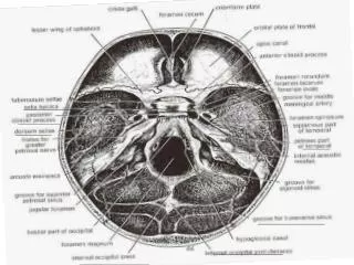
Meninges: Vascular Supply • Anterior meningeal arteries: From ethmoidal arteries and internal carotid. • Middle meningeal artery: From maxillary artery. • Emissary veins: Between dural venous sinuses and external veins. • Diploic veins. • Cerebral veins.
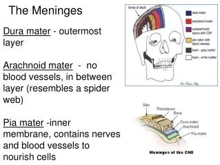
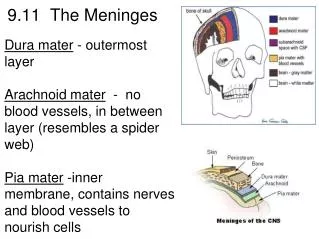
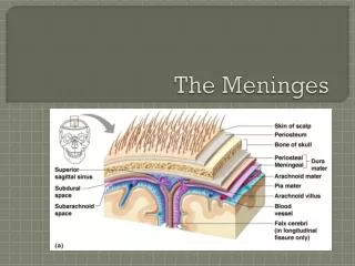
Meninges: Dura Mater • Tough outer connective tissue layer that forms sac around brain. • No epidural space in skull. • Encloses dural venous sinuses.
Meninges: Dura Mater • Reflections: Falx cerebri: Midline fold of dura mater extending between two cerebral hemispheres. Tentorium cerebelli: Dural fold located between cerebellum and occipital lobes of cerebral hemispheres.
Meninges: Dura Mater • Reflections: Falx cerebelli: Dural fold between two cerebellar hemispheres. Diaphragma sellae Dural fold over hypophyseal fossa.
Meninges: Dura Mater • Dural venous sinuses: Superior sagittal sinus: Lies along superior margin of falx cerebri. Inferior sagittal sinus: Lies along inferior margin of falx cerebri.
Meninges: Dura Mater • Dural venous sinuses: Straight sinus: Lies at intersection of falx cerebri and tentorium cerebelli. Confluence of sinuses: Common confluence of superior sagittal sinus and straight sinus.
Meninges: Dura Mater • Dural venous sinuses: Transverse: Begins at confluence of sinuses. Extends along edges of tentorium cerebelli. Right receives blood from superior sagittal sinus. Left receives blood from straight sinus.
Meninges: Dura Mater • Dural venous sinuses: Sigmoid: Continuation of straight sinus. “S”-shaped. Ends at jugular foramen: Joins internal jugular vein.
Meninges: Pia Mater • Innermost layer. • Closely applied to surface of brain. • Dips into fissures and sulci. • Forms sheath around blood vessels as they penetrate surface of brain.
Meninges: Arachnoid • Intermediate layer. • Attached to dura mater and pia mater: Separated from pia mater via: Subarachnoid space: Contains cerebrospinal fluid.
Meninges: Arachnoid • With many arachnoid villi: For reabsorption of cerebrospinal fluid. Arachnoid granulations may pit surrounding bone: Fovea granulares.
Meninges: Arachnoid • Subarachnoid cisterns: Choroid plexuses. Flow of CSF: Arachnoid villi. Arachnoid granulations. • Hydrocephalous: Obstructive. Communicating.