Download
1 / 66
700 likes | 980 Views
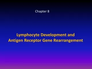
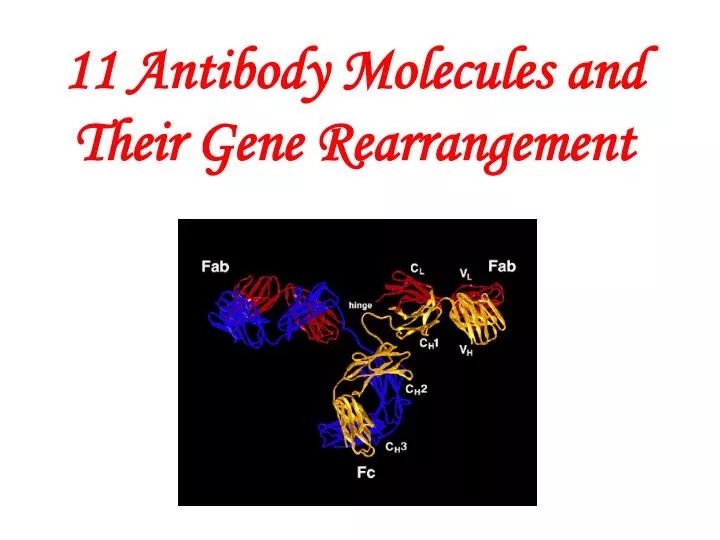
11 Antibody Molecules and Their Gene Rearrangement. Master the concepts of Ig and Ab; Master the relationship between structure and functions of Ab; Master properties and biological activities of five classes of Igs. Emil von Behring, 1901, antitoxins.

E N D
Master the concepts of Ig and Ab; Master the relationship between structure and functions of Ab; Master properties and biological activities of five classes of Igs.
Emil von Behring, 1901, antitoxins Paul Ehrlich , 1908, production of antibody Georeges Kohler and Cesar Milstein, 1984, monoclonal antibody Gerald Edelman and Rodney Porter, 1972, structure of antibody Susumu Tonegama,1987, structure of Ig gene Nobel Prize winners
Emil von Behring (1845-1917) Emil von Behring, 1901, antitoxins
1. Antibody (Ab) 2. Immunoglobulins (Ig) 3. The relationship between Ab & Ig:
Antibody (Ab) A globulin which is produced by plasma cell as a result of the introduction of an antigen and which has the ability to combine with the antigen that stimulated its production. Secretion Differatiation
Immunoglobulins (Ig) Globulins composed of H and L chains, or globulins function as antibody. Immunoglobulins Antibody-containing serum is place in an electrical field Antibodies migrated with the globular proteins.
Immune sera Normal sera Figure 1 Electrophoretic separation of serum proteins
The relationship between Ab & Ig All antibodies are immunoglobulins, but it is not certain that all immunoglobulins have antibody function.
Distribution of immunoglobulins Immunoglobulins (antibody) molecules are found in serum (account for approximately 20% of the total plasma protein ), in extravascular fluids, in exocrine secretions, and on the surface of some lymphocytes.
IgM B Cell Expressed on the surfaces of resting B lymphocytes, server as RECEPTORS that can detect and distinguish among the vast array of potential antigens present in the environment.
Basic structure of Antibody Molecules * A four polypeptide chains: two identical light chains two identical heavy chains , held by disulfide bonds. ** Y-shape structure, symmetric. *** –NH2 terminal, -COOH terminal. **** variable & constant regions. ***** domains
Heavy chain (H) and light chain H chain ① Composed of about 500 aa, oligosaccharide(+) ② Class : heavy chain , , , , . immunoglobulin(Ig) IgA, IgG, IgM IgD, IgE L chain ① Composed of about 214 aa, oligosaccharide(-) ② Type: ,
V region ①N-terminal1/2L+1/4(1/5)H; VL, VH
V region N-terminal 1/2L+1/4(1/5)H; Constant region (C-terminal 1/2L+3/4(4/5)H VL VH CL CH1 CH2 CH3 (CH4)
CH2 CH3 CH4 Hinge region NH3+ VH VL Properties: 1) Flexible 2) Rich in proline Function: 1) Facilitating the interaction between Ag and Ab 2) Facilitating complement fixation CH1 CL Hinge region COO–
IgG Molecule Conformational Changes Induced by Antigen Binding POSTBINDING PREBINDING Fab CH1 Exposed C1q-binding site Barricaded C1q-binding site CH2 Fc (IgM CH3,IgG CH2)
Figure 3-3 part 1 of 2 Enzymatically generated Ab fragments (1) Papain(木瓜蛋白酶): Fab (Ag-binding Fragment ) Fc (Crystallizable Fragment ) : complement fixation, FcR
Figure 3-3 part 2 of 2 (胃蛋白酶) (2) Pepsin(胃蛋白酶) ( Fab’ ) 2 pFc ( peptides of Fc )
Tertiary structure of antibody molecules Hypervarible region (HVR) (complimentarity determining region,CDR) : formation of the Ag binding site
CDR (complimentarity determining region,) Light chain
antibody antigen-antibody complex: antigen purple : HV CDR ( in both the ribbon and ball and stick views) green : antigen HV sequences contact the antigen.
V region: Ag binding • Neutralizing toxin & virus • Agglutination microbes, • Prevention adhesion
C region: Fixation of complement Binding cells Opsonization Mediating ADCC
Opsonization (调理作用): The process of attaching opsonins (调理素), such as IgG or complement fragments, to microbial surfaces to target the microbes for phagocytosis.
Recognition of microbes by neutrophils and macrophages complement antibody Surface receptor on macrophage CD11b/CD18 CD16 (FcR III) IL-2 Lactoferrin CD25 CD71 B7-2 CD28 CD64 (Fc R I) CD35 (CR1) CD32 (Fc R II) antibody complement antibody
FcgR and Complement Receptors Cooperate To Induce Greater Phagocytosis
(2) ADCC FcgRIII CD16Antibody Marks Target Cells For NK Cell Attack (ADCC)
IgG (Immunoglobulin G) 7.4.1 11 interchain disulfide bonds.
Four Subclasses of Human IgG General structure of the four subclasses of human IgG, which differ in the number and arrangement of the interchain disulfide bonds linking the heavy chains. A notable feature of human IgG3 is its 11 interchain disulfide bonds. IgG1 IgG2 IgG3 IgG4
Properties (A) IgG is the major Ig in serum - 75% (B) The longest half life (t ½ =23days) (C) IgG is the major Ig in extravascular spaces (D) Placental transfer (E) Fixation complement – (F) Binding to cells –Opsonization mediating ADCC Immunity is transferred from mother to fetus through placental transfer of IgG.
Iga Igb Iga Igb IgM (Immunoglobulin M) (1) Structure Secreted IgM (sIgM): pentamer Membrane-bound IgM (mIgM): monomer J chain
Joining chain (1)Chemical nature: polypeptide chain secreted by plasma cell Secrete piece J CHAIN Joining chain IgM IgA (2) Presence: polymeric Igs such as IgM (pentamer), sIgA (dimer).
(2) Properties (A) The first Ig made by fetus and B cells (B) Fixation complement –classical pathway (C) The largest size of molecule (D) Natural blood type antibody (E) Binding to cells – Opsonization.
IgA (Immunoglobulin A) (1) Structure