Download
1 / 18
180 likes | 243 Views
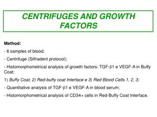
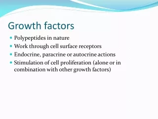
Explore the influence of Medifuge-Silfradent on growth factors in blood samples through histomorphometrical and quantitative analysis, revealing higher expressions of VEGF-A and TGF-β1. Enzymatic assays confirm decreased serum concentrations of these growth factors compared to control centrifuge treatments. Immunohistochemical analysis indicates a greater presence of CD34+ stem cells in the Red-Buffy Coat Interface following Medifuge-Silfradent treatment.

E N D
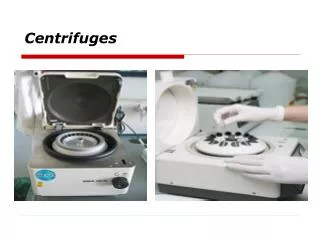
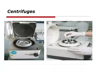
CENTRIFUGES AND GROWTH FACTORS • Method: • 6 samples of blood; • Centrifuge (Silfradent protocol); • Histomorphometrical analysis of growth factors: TGF-1 e VEGF-A in Buffy Coat: • 1) Buffy Coat, 2) Red-buffy coat Interface e 3) Red Blood Cells 1, 2, 3; • Quantitative analysis of TGF-1 e VEGF-A in blood serum; • - Histomorphometrical analysis of CD34+ cells in Red-Buffy Coat Interface.
MEDIFUGE- SILFRADENT CONTROL CENTRIFUGE SERUM BUFFY COAT RED BLOOD CELLS Buffy Coat Red- Buffy Coat Interface Red Blood 1 Red Blood 2 Red Blood 3
* * P < 0,05 vs control centrifuge VEGF-A serum VEGF-A serum concentration is lower in the samples treated with Medifuge-Silfradent compared with control centrifuge.
* * * * * *P < 0,05 vs control centrifuge VEGF-A expression The histomorphometrical analysis showed a greater expression of VEGF-A in the samples treated with Medifuge-Silfradent compared with control centrifuge.
* * P < 0,05 vs control centrifuge VEGF-A Buffy Coat CONTROL Histomorphometrical analysis (IOD) of VEGF-A showed a greater expression of this growth factor in the Buffy Coat of samples treated with Medifuge-Silfradent compared with control centrifuge. MEDIFUGE
* * P< 0,05 vs control centrifuge VEGF-A Red – Buffy Coat Interface CONTROL Histomorphometrical analysis (IOD) of VEGF-A showed a greater expression of this growth factor in the Buffy Coat of samples treated with Medifuge-Silfradent compared with control centrifuge. MEDIFUGE
* * P< 0,05 vs control centrifuge VEGF-A Red Blood 1 CONTROL Histomorphometrical analysis (IOD) of VEGF-A showed a greater expression of this growth factor in the Red Blood 1 of samples treated with Medifuge-Silfradent compared with control centrifuge. MEDIFUGE
* * P< 0,05 vs control centrifuge VEGF-A Red Blood 2 CONTROL Histomorphometrical analysis (IOD) of VEGF-A showed a greater expression of this growth factor in the Red Blood 2 of samples treated with Medifuge-Silfradent compared with control centrifuge. MEDIFUGE
* * P< 0,05 vs control centrifuge VEGF-A Red Blood 3 CONTROL Histomorphometrical analysis (IOD) of VEGF-A showed a greater expression of this growth factor in the Red Blood 3 of samples treated with Medifuge-Silfradent with control centrifuge. MEDIFUGE
* * P< 0,05 vs control centrifuge TGF-β1 Serum TGF-β1serum concentration is lower in samples treated with Medifuge-Silfradent compared with control centrifuge.
TGF-β1 expression * * * * * * P < 0,05 vs control centrifuge The histomorphometrical analysis showed a greater expression of TGF-β1in samples treated with Medifuge-Silfradent compared with control centrifuge.
* * P < 0,05 vs control centrifuge TGF-β1Buffy Coat CONTROL Histomorphometrical analysis (IOD) of TGF-β1 showed a greater expression of this growth factor in the Buffy Coat of samples treated with Medifuge-Silfradent compared with control centrifuge. MEDIFUGE
* * P < 0,05 vs control centrifuge TGF-β1 Red – Buffy Coat Interface CONTROL Histomorphometrical analysis (IOD) of TGF-β1 showed a greater expression of this growth factor in the Red-Buffy Coat of samples treated with Medifuge-Silfradent compared with control centrifuge. MEDIFUGE
* * P < 0,05 vs control centrifuge TGF-β1 Red Blood 1 CONTROL Histomorphometrical analysis (IOD) of TGF-β1 showed a greater expression of this growth factor in the Red Blood 1 of samples treated with Medifuge-Silfradent compared with control centrifuge. MEDIFUGE
* * P < 0,05 vs control centrifuge TGF-β1 Red Blood 2 CONTROL Histomorphometrical analysis (IOD) of TGF-β1 showed a greater expression of this growth factor in the Red Blood 2 of samples treated with Medifuge-Silfradent compared with control centrifuge. MEDIFUGE
* * P < 0,05 vs control centrifuge TGF-β1 Red Blood 3 CONTROL Histomorphometrical analysis (IOD) of TGF-β1 showed a greater expression of this growth factor in the Red Blood 3 of samples treated with Medifuge-Silfradent compared with control centrifuge. MEDIFUGE
VEGF-A and TGF-β1 results • The histomorphometric analysis (IOD) of blood growth factors TGF-1 and VEGF-A showed a greater expression of these growth factors in the samples treated with Medifuge-Silfradent compared with control centrifuge. • Enzymatic analysis of VEGF-A e TGF-1 has confirmed our previous results showing a significant difference of both growth factors serum concentration: in particular, we found a lower serum concentration of both growth factors in the samples treated with Medifuge-Silfradent compared with control centrifuge. Bar 10 μm
CD 34 Stem cells Red- Buffy Coat Interface CONTROL MEDIFUGE • - The immunohistochemical analysis of the Red-Buffy Coat Interface showed a greater number of white cells in samples treated with Medifuge-Silfradent centrifuge with control centrifuge, as suggest by our previous analysis. • - The immunohistochemical analysis of cells CD34+ expressed in the Red-Buffy Coat Interface showed a number of cells CD34+ four time greater in the samples trated with Medifuge-Silfradent centrifuge compared with control centrifuge.