Download
1 / 29
320 likes | 604 Views
The Nose and Para nasal Sinuses 241 RTS. Prepared by : Dr. Taghrid M. Abdallah . Dr.Taghrid@inaya.edu.sa. EXTERNAL NOSE. Projects from the face Vary in size and shape due to the differences in nasal cartilage Parts : 1] Root – superior angle 2] Apex – tip

E N D
The Nose and Para nasal Sinuses241 RTS Prepared by: Dr. Taghrid M. Abdallah. Dr.Taghrid@inaya.edu.sa
EXTERNAL NOSE • Projects from the face • Vary in size and shape due to the differences in nasal cartilage • Parts: 1] Root – superior angle 2] Apex – tip 3] Nares (nostrils): 2 pyriform openings 4] Alae – lateral boundaries 5] Nasal septum – middle struc w/c separates 2 nares 6] Vestibule – w/ stiff hairs
Skeletal part • Nasal bones • Frontal proc of maxillae • Nasal part of frontal bone Cartilaginous Part • 2 Lateral cartilage • 2 Alar cartilage • 1 Septal cartilage
Nasal Septum • Divides the chamber of the nose into 2 nasal cavities: • Components: 1] Perpendicular plate of ethmoid- forms superior part of the nasal septum 2] Vomer- thin flat bone 3] Septal cartilage- articulates w/ bony septum
Nasal Cavity • Open posteriorly into the nasopharynx thru the choanae • Mucosa lines the nasal cavity except for vestibule • Nasal mucosa 1] Olfactory area • superior 1/3 • contains organ of smell 2] Respiratory area • inferior 2/3 • where air is warmed and moistened before it goes to the respiratory tract.
Linings of nasal cavity • Vestibule* (just above nostrils) • Lined with skin containing sebaceous and sweat glands and nose hairs • Filters large particulars (insects, lint, etc.) • The remainder of nasal cavity: 2 types of mucous membrane • Small patch of olfactory mucosa near roof (cribriform plate) • Respiratory mucosa: lines most of the cavity
Respiratory Mucosa • Pseudostratified ciliated columnar epithelium • Scattered goblet cells • Underlying connective tissue lamina propria • Mucous cells – secrete mucous • Serous cells – secrete watery fluid with digestive enzymes, e.g. lysozyme • Together all these produce a quart/day • Dead junk is swallowed
Nasal Concha • 3 scroll-shaped elevations • Superior,middle,inferior • Divide the nasal cavity into 4 passages 1] Sphenoethmoidal recess 2] Superior meatus 3] Middle meatus 4] Inferior meatus
Nasal Passages • Sphenoethmoidal recess - sphenoidal sinus • Superior meatus -posterior ethmoidal sinus • Middle meatus -anterior/middle ethmoidal sinus -frontal sinus -maxillary sinus • Inferior meatus -nasolacrimal duct
Vasculature • Arterial Supply: Branches of: 1.Sphenopalatine artery 2.Anterior and posterior ethmoidal artery. 3.Greater palatine artery. 4.Superior labial artery. 5.Lateral nasal branches of facial artery. >Kiesselbach’s area- anterior, where all arteries anastomose, where nose bleeding occurs
Vasculature • Venous drainage - Sphenopalatine vein. - Facial vein. - Ophthalmic vein.
Nerve Supply • Nasal septum • Nasopalatine n • Greater palatine n • Anterior ethmoidal n • Posterior ethmoidal n • Smell • Olfactory n • Olfactory bulb - forebrain
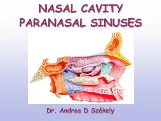
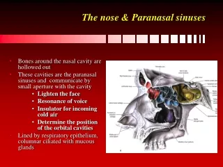
PARANASAL SINUSES • Air-filled extensions of the respiratory part of the nasal cavity into the ff cranial bones: • FRONTAL • ETHMOID • SPHENOID • MAXILLARY
Paranasal sinuses • Frontal, sphenoid, ethmoid and maxillary bones • Open into nasal cavity • Lined by same mucosa as nasal cavity and perform same functions • Also lighten the skull • Can get infected: sinusitis
FRONTAL SINUS • Between outer and inner tables of frontal bone • Detectable by 7 yo • Drains thru frontonasal duct into infundibulum w/c opens into semilunar hiatus of middle meatus • NS: br of supraorbital n
ETHMOIDAL SINUS • Comprise several cavities-ethmoidal cells • At ethmoid bone • Parts: • Anterior –drain into middle meatus • Middle – form ethmoidalbulla,drain into middle meatus • Posterior - drain into superior meatus • NS: Nasociliary n[CN V1]
Sphenoidal Sinus • Unevenly divided • Body of sphenoid • Separated by thin plates of bone from optic nerve and chiasma,ICA, pit’y gl • Opens into sphenoethmoidal recess • NS: posterior ethmoidal nerve
Maxillary Sinus • Largest • Pyramidal shaped • Parts: Apex- into zygomatic bone Base- part of nasal cavity Roof- formed by floor of orbit Floor- formed by alveolar part of maxilla Drains via maxillary ostium into middle meatus NS: Superior alveolar nerves
Thank you • Best Wishes