Download
1 / 33
360 likes | 477 Views
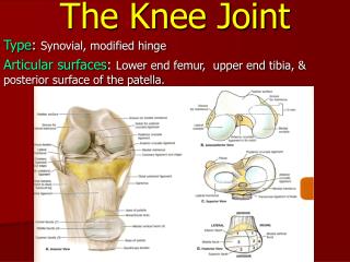
The Knee Joint. The Knee Joint. Knee joint largest joint in body very complex primarily a hinge joint. Bones. Enlarged femoral condyles articulate on enlarged tibial condyles Medial & lateral tibial condyles (medial & lateral tibial plateaus) - receptacles for femoral condyles

E N D
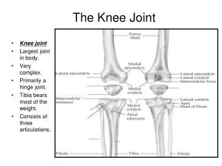
The Knee Joint • Knee joint • largest joint in body • very complex • primarily a hinge joint
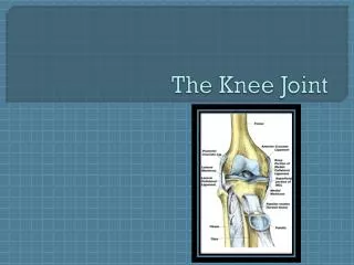
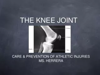
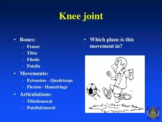
Bones • Enlarged femoral condyles articulate on enlarged tibial condyles • Medial & lateral tibial condyles (medial & lateral tibial plateaus) - receptacles for femoral condyles • Tibia – medial • bears most of weight
Bones • Fibula - lateral • serves as the attachment sight for knee joint structures • does not articulate with femur or patella • not part of knee joint
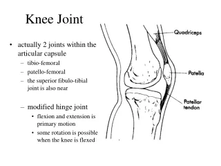
Bones • Patella • sesamoid (floating) bone • imbedded in quadriceps & patellar tendon
Bones • Key bony landmarks • Tibial tuberosity • Gerdy’s tubercle • Medial & lateral femoral condyles • Medial tibial plateau • Lateral tibial plateau • Head of fibula
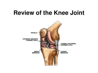
Joints • Ligaments provide static stability • Quadriceps & hamstrings contractions produce dynamic stability • Articular cartilage surfaces on femur & tibia • Menisci form cushions between bones • attached to tibia • deepen tibial fossa • enhance stability
Joints • Medial meniscus forms receptacle for medial femoral condyle, Lateral meniscus receives lateral femoral condyle • Thicker on outside border & taper down very thin to inside border • Can slip about slightly, but held in place by various small ligaments • Medial meniscus - larger & more open C appearance • Lateral meniscus - closed C configuration
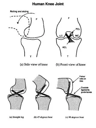
Joints • Anterior & posterior cruciate ligaments • cross within knee between tibia & femur • vital in respectively maintaining anterior & posterior stability, as well as rotatory stability • Anterior cruciate ligament (ACL) injuries • one of most common serious injuries to knee • mechanism often involves noncontact rotary forces associated with planting & cutting, hyperextension, or by violent quadriceps contraction which pulls tibia forward on femur
Joints • Posterior cruciate ligament (PCL) injuries • not often injured • mechanism of direct contact with an opponent or playing surface • Lateral collateral ligament (LCL) • infrequently injured
Joints • Medial collateral ligament (MCL) • maintains medial stability by resisting valgus forces or preventing knee from being abducted • injuries occur commonly, particularly in contact or collision sports
Joints • Extends to 180 degrees (0 degrees of flexion) • Hyperextension of 10 degrees or > not uncommon • Flexion occurs to about 140 degrees • With knee flexed 30 degrees or > • internal rotation 30 degrees occurs • external rotation 45 degrees occurs
Joints • Knee “screws home” to fully extend due to the shape of medial femoral condyle • As knee approaches full extension tibia must externally rotate approximately 10 degrees to achieve proper alignment of tibial & femoral condyles • In full extension • close congruency of articular surfaces • no appreciable rotation of knee • During initial flexion from full extension • knee “unlocks” by tibia rotating internally, to a degree, from its externally rotated position to achieve flexion
Movements • Flexion • bending or decreasing angle between femur & leg, characterized by heel moving toward buttocks • Extension • straightening or increasing angle between femur & lower leg
Movements • External rotation • rotary movement of leg laterally away from midline • Internal rotation • rotary movement of lower leg medially toward midline • Neither will occur unless flexed 20-30 degrees or more
Muscles • Quadriceps muscle group • extends knee • located in anterior compartment of thigh • consists of 4 muscles • rectus femoris • vastus lateralis • vastus intermedius • vastus medialis
Muscles • Hamstring muscle group • responsible for knee flexion • located in posterior compartment of thigh • consists of 3 muscles • semitendinosus - medial, internal rotator • semimembranosus - medial, internal rotator • biceps femoris - lateral, external rotator • Popliteus assist medial hamstrings in knee internal rotation
Muscles Knee joint muscles location • Anterior - primarily knee extension • Rectus femoris • Vastus medialis • Vastus intermedius • Vastus lateralis
Muscles Knee joint muscles location • Posterior - primarily knee flexion • Biceps femoris • Semimembranosus • Semitendinosus • Sartorius • Gracilis • Popliteus • Gastrocnemius
Rectus Femoris Muscle Flexion of hip Extension of knee Anterior pelvic rotation
Vastus Lateralis Muscle Extension of knee
Vastus Intermedius Muscle Extension of knee
Vastus Medialis Muscle Extension of knee
Hamstring Muscles • Hamstring muscle group • Semitendinosus • Biceps femoris • Semimembranosus
Hamstring Muscles • Hamstring muscle strains very common • “Running muscles” function in acceleration • Antagonists to quadriceps muscles at knee • Named for cordlike attachments at knee • All originate on ischial tuberosity of pelvis • Semitendinosus inserts on anteromedial tibia • Semimembranosus inserts on posteromedial tibia • Biceps femoris inserts on lateral tibial condyle & head of fibula
Semitendinosus Muscle Flexion of knee Extension of hip Internal rotation of hip Internal rotation of flexed knee Posterior pelvic rotation
Semimembranosus Muscle Flexion of knee Extension of hip Internal rotation of hip Internal rotation of flexed knee Posterior pelvic rotation
Biceps Femoris Muscle Flexion of knee Extension of hip External rotation of hip External rotation of flexed knee Posterior pelvic rotation
Popliteus Muscle Flexion of knee Internal rotation of flexed knee
Knee Extension • Agonists • Rectus Femoris • Vastus Lateralis • Vastus Intermedius • Vastus Medialis
Knee Flexion • Agonists • Biceps Femoris (Long & Short Head) • Semitendinosus • Semimembranosus
Knee Internal Rotation • Agonists • Semitendinosus • Semimembranosus • Popliteus
Knee External Rotation • Agonists • Biceps Femoris