Download

1 / 55
560 likes | 2.87k Views
THE ITCHY VULVA LICHEN SCLEROSUS (LS). L.J. Margesson , MD + G. Davis, MD. Introduction Lichen Sclerosus. A common chronic vulvar disease An inflammatory skin condition Prevalence 1 in 300 to 1 in 1,000

E N D
THE ITCHY VULVALICHEN SCLEROSUS (LS) L.J. Margesson, MD + G. Davis, MD
Introduction Lichen Sclerosus A common chronic vulvar disease An inflammatory skin condition Prevalence 1 in 300 to 1 in 1,000 Most commonly found in middle-age women, but it can be seen in very young children and the elderly
Etiology of LS Unknown Multifactorial - genetic - autoimmune - environmental factors NOTE: Often associated with autoimmune conditions, e.g. thyroid disease, vitiligo, etc. Familial cases have been reported
Clinical Findings LS SYMPTOMS: Most common - pruritus - can be severe, intolerable Can have soreness and burning Often asymptomatic Scratching results in open areas causing dysuria, pain, dyspareunia, etc.
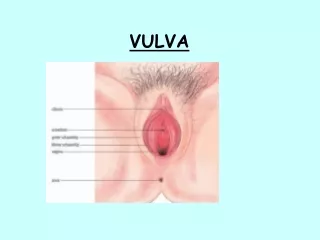
Clinical Findings LS PHYSICAL EXAMINATION: Ivory white papules or confluent plaques Cellophane-like sheen to surface Patchy or generalized - anywhere vulva, perineum, perianal Disease is not in the vagina
Clinical Findings LS PHYSICAL EXAMINATION Secondary changes - scratches, purpura, erosion - crusting, thickening (lichenification) - scarring with loss of normal architecture; fusion of labia minora -phimosis -introital stenosis NOTE: LS can be associated with squamouscell carcinoma of the vulva. Any open, raw, non-healing lesions must be assessed and biopsied.
Case History LS 35-year-old woman with vulvar itching for 2½ years - treated repeatedly for yeast infections - topical products burn her - was told the area looked thinned and pale; questionable pre-menopause
Case LS PHYSICAL EXAMINATION: 35-year-old woman No other skin rashes Bilaterally symmetrical white rash Extends from supra-clitoral to perianal area Slight scarring around the clitoris
Diagnosis LS Clinical Pattern Biopsy
Histopathology LS Thinned epidermis +/- hyperkeratosis In reticular dermis, a band of homogenized collagen Lymphocytic infiltrate under the band In individuals who scratch chronically, histologic changes of squamous cell hyperplasia can be seen in addition to LS changes
Treatment of LS I Thorough assessment Biopsy to confirm diagnosis Stop irritants Education Superpotent steroid ointment clobetasol 0.05% ointment bid x 1 month, then qhs x 2 months. Apply in a thin film. Maintenance dose is 1 to 3 times per week indefinitely Treat secondary infection, particularly yeast
Treatment of LS Stop scratching Consider sedation, if necessary If very thick, consider intralesional triamcinolone acetonide If not responding, reassess, rebiopsy to R/O SCC, R/O contact dermatitis Set up long-term followup
References LS Dalziel K, Shaw S. Lichen sclerosus.BMJ. 2010;340:c731. Lynch PJ, Moyal-Barrocco M, Bogliatto F, Micheletti L, Scurry J. 2006 ISSVD classification of vulvardermatoses: pathologic subsets and their clinical correlates. Journal of Reproductive Medicine. 2007;52(1):3-9.
References LS (cont’d) Lynch, PJ. 2006 International Society for the Study of Vulvovaginal Disease Classification of VulvarDermatoses: A Synopsis. J Low Gen Tract Dis 2007;11(1):1-2. Saunders NA, Haefner HK. Vulvar lichen sclerosus in the elderly: pathophysiology and treatment update. Drugs & Aging. 22009;6(10):803-12.
References LS (cont’d) Smith SD, Fischer G. Paediatricvulval lichen sclerosusAustralasian Journal of Dermatology. 2009;50(4):243-8. van der Avoort IA, Tiemes DE, van Rossum MM, van der Vleuten CJ, Massuger LF, de Hullu JA. Lichen sclerosus: treatment and follow-up at the departments of gynaecology and dermatology. Journal of Lower Genital Tract Disease. 2010;14(2):118-23.
THE ITCHY VULVASQUAMOUS CELL HYPERPLASIALICHEN SIMPLEX CHRONICUS (LSC) L. J. Margesson, MD + G. Davis, MD
Squamous Cell Hyperplasia Is Lichen Simplex Chronicus
Introduction - LSC With this condition there is chronic, intense itching that results in repetitive scratching and rubbing The skin responds by thickening (lichenification). The thickening of the skin is caused by the scratching An itch-scratch-itch cycle starts and perpetuates the problem
Etiology LSC I LSC develops in several itchy skin conditions: Atopic dermatitis (eczema) Contact dermatitis Lichen sclerosus Contact dermatitis can start this condition or be the main long-term promoting factor
Etiology LSC II Scratching and rubbing damage the skin so it loses its protective coating / barrier Result is: susceptibility to infection ease of irritation more itching
Clinical Findings LSC SYMPTOMS: Relentless pruritus Years of itch Itch eventually developing into burning pain Worse with heat, stress, periods and tight synthetic clothing Wake up at night scratching
Clinical Findings LSC PHYSICAL EXAMINATION: Thick, lichenified skin so labia are enlarged, rugose +/- edematous Bilateral or unilateral Localized or generalized Color - variably pink, red, violaceous to ruddy brown; often a white appearance will be present when there is a thick keratin layer deposited on the surface of the epithelium Secondary changes - erosions, ulcers, oozing, fissuring, honey-colored or serosanguineous crusting
CASE HISTORY: LSC 26-year-old woman with intense vulvar pruritus for 2½ years Wakes up at night scratching Nothing helps Most creams burn her Has allergic rhinitis and asthma
Case LSC PHYSICAL EXAMINATION: A healthy, 26-year-old woman No other skin changes On the inner labia majora, interlabial sulcus and perineum is a bilateral, symmetrical, pink, lichenified eruption Some excoriations seen
DIAGNOSIS LSC Clinical Do biopsy to R/O underlying condition Culture for secondary infection - yeast and bacteria Consider patch testing
Histopathology LSC Hyperkeratosis Acanthosis Lengthened broad rete ridges Chronic inflammatory infiltrate
Treatment LSC I Stop the itch-scratch-itch cycle Sitz baths and soaks, no irritants Reduce inflammation with superpotent steroids, i.e., clobetasol or halobetasol ointment - bid for two weeks, - once a day for two weeks and - M / W / F for two weeks Intralesional steroids, if severe
Treatment LSC I Intralesional steroids -5 mg of triamcinolone suspension in 2 ml of saline subcutaneously -Use a 3-inch, 22-gauge spinal needle inserted in the lower mons pubis and then passed down to the perineum -As the needle is withdrawn, the solution is slowly injected and then massaged into the tissue Systemic steroids are seldom needed
Treatment LSC Control infection - cefadroxil 500 mg bid for 7 days, fluconazole 150 mg repeated one week later Sedation: doxepin or hydroxyzine 25-10 mg qhs, SSRI like fluoxetine 20 mg q am
References LSC O'Connell TX, Nathan LS, Satmary WA, Goldstein AT. Non-neoplastic epithelial disorders of the vulva. American Family Physician. 2008;77(3):321-6. Burrows LJ, Shaw HA, Goldstein AT. The vulvardermatoses. Journal of Sexual Medicine. 2008;5(2):276-83.
THE ITCHY VULVALICHEN PLANUS (LP) L. J. Margesson, MD + G. Davis, MD
Introduction LP An inflammatory, mucocutaneous eruption With a distinctive pattern on: - skin, scalp, nails - mucous membranes oral, genital, esophageal, etc.
Etiology LP Unknown ? Autoimmune triggered by exogenous antigens, possibly - viral - bacterial (superantigen) - chemical - drug - trauma
Clinical Findings LP SYMPTOMS Most often there is irritation with burning and soreness Can be very itchy, and scratching flares it With scratching, the vulva gets very thick and scarred Stretching of scarring causes dyspareunia Symptoms depend on extent of disease - e.g. when vagina is involved with erosions, there is discharge, burning, etc.
Clinical LP PHYSICAL EXAMINATION Variable patterns Lacey, reticulated pattern on the labia, vulvar trigone, perineum or perianal area with or without scarring and erosions. Small reddish papules may be seen
Clinical LP PHYSICAL EXAMINATION Thick, white, indurated plaques anywhere from clitoris to anus – as in lichen sclerosus Secondary changes: excoriations, crusting, scarring If ulcerative: vulva - glazed erythema, erosions, ulcers vagina - synechiae, scarring, stenosis, telescoping, bleeding, discharge
Diagnosis LP Look at rest of skin and mucous membranes Look in the mouth Biopsy - regular histopathology (H&E) - immunofluorescence Stop topical steroids for 1-2 weeks before biopsy
Case LP A 42-year-old woman presented with severe itching and burning on her vulva for 18 months Topical treatment for yeast and herpes simplex burned her Topical cortisone-no help Initial dyspareunia now apareunia
Case LP PHYSICAL EXAMINATION Lacey white pattern of involvement in interlabial sulci Peri-clitoral whitish plaque Ulceration of clitoris and interlabial sulci Early scarring of the clitoris
Histopathology LP Hyperkeratosis Prominent granular layer Irregular acanthosis and sawtooth pattern Destruction of basal layer Band-like inflammatory infiltrate
Treatment LP Challenging - no single agent is universally effective Stop irritation and trauma Treat infection Restore barrier function with Sitz bath or tub bath 1- 2 times a day Reduce inflammation with topical superpotent corticosteroids clobetasol 0.05% ointment 1- 2 times a day
Treatment LP For the vagina - hydrocortisone acetate foam (80 mg) at night or in a compounded suppository 100 mg For localized disease consider intralesional triamcinolone
Treatment LP Severe Lichen Planus control: Prednisone 1 - 1.5 mg per kg per day for 2 weeks and tapering over 2-4 months or IM triamcinolone 1 mg per kg q 4 weeks for maximum three injections not to exceed 80 mg per month Add cyclosporine 4 mg per kg per day and continue until the patient is clear, then wean onto Plaquenil 200 mg bid and / or hydrocortisone acetate vaginally Other drugs to consider: doxycycline, metronidazole, acitretin, methotrexate, azathioprine and tacrolimus
Treatment LP Tacrolimus 0.03% and 0.1% ointment can be used topically but the Protopic brand can sting and must be used only on areas under control with topical steroids - used as steroid sparers Custom formulas are available for suppository and intravaginal creams and ointments
Prognosis LP 38% complete resolution 30% significant resolution 32% ongoing problems