Download
1 / 94
940 likes | 959 Views
Discover the fascinating journey of early microscopes from simple to compound and delve into the advanced world of modern electron microscopes. Learn about the Cell Theory proposed by prominent scientists and the importance of cell size and shape.

E N D
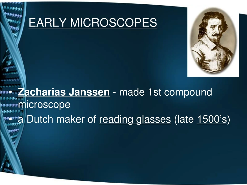
EARLY MICROSCOPES • Zacharias Janssen - made 1st compound microscope • a Dutch maker of reading glasses (late 1500’s)
Leeuwenhoek • made a simple microscope (mid 1600’s) • magnified 270X • Early microscope lenses made images larger but the image was not clear
Leeuwenhoek's microscope A) a screw for adjusting the height of the object being examined B) a metal plate serving as the body C) a skewer to impale the object and rotate it D) the lens itself, which was spherical
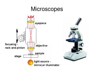
MODERN MICROSCOPES • A microscope is simple or compound depending on how many lenses it contains • A lens makes an enlarged image & directs light towards you eye
A simple microscope has one lens • Similar to a magnifying glass • Magnificationis the change in apparent size produced by a microscope
COMPOUND MICROSCOPE • A compound microscope has multiple lenses • (eyepiece & objective lenses)
Microscope • Magnification increases the size of an object. • As the magnification increases your field of view decreases. • Resolution increases the ability to see details of the specimen
TOTAL MAGNIFICATION • Powers of theeyepiece (10X) multiplied by objective lenses determine total magnification.
STEREOMICROSCOPE • creates a 3D image
ELECTRON MICROSCOPES • More powerful; some can magnify up to 1,000,000X • Use a magnetic field in a vacuum to bend beams of electrons • Images must be photographed or produced electronically
ScanningElectron Microscope (SEM) Electron microscope image of a spider Electron microscope image of a fly foot • produces realistic 3D image • only the surface of specimen can be observed
SEM Images Dust Mite Pollen
SEM Sickle Cell Virus
TransmissionElectron Microscope (TEM) • produces 2D image of thinly sliced specimen • detailed cell parts (only inside a cell) can be observed
ScanningTunneling Microscope (STM) • able to show arrangement of atoms
Scanning Tunneling Microscope(STM) • Device for studying and imaging individual atoms on the surfaces of materials. The instrument was invented in the early 1980s by Gerd Binnig and Heinrich Rohrer, who were awarded the 1986 Nobel prize in physics for their work.
The underlying principle of the STM is the tunneling of electrons between the sharp tip of a probe and the surface of the sample under study. The flow of electrons is extremely sensitive to the distance between the tip and the sample. As the tip is swept over the surface the height of the tip is continually adjusted so as to keep the flow of electrons constant. A map of the "bumps" on the surface is then obtained by accurately recording the height fluctuations of the tip. • The STM was used in 2004 to measure the charges of individual atoms, and in 2010 researchers used a modified STM to observe the magnetism, or spin, of atoms on the nanosecond timescale.
CELL THEORY • A theory resulting from many scientists’ observations & conclusions
CELL THEORY1. The basic unit of life is the cell. (Hooke) • In 1665, an English scientist named Robert Hooke made an improved microscope and viewed thin slices of cork viewing plant cell walls • Hooke named what he saw "cells"
CELL THEORY2. All living things are made of 1 or more cells. • Matthias Schleiden(botanist studying plants) • Theodore Schwann(zoologist studying animals) stated that all living things were made of cells Schwann Schleiden
CELL THEORY3. All cells divide & come from oldcells. (Virchow) Virchow
Cell Theory • Hooke and Leeuwenhooke recorded all that they saw. • Schleiden, Swann, & Virchow further studied cells and proposed The Cell Theory. Page 55.
Cell Theory • All living organisms are composed of one or more cells. • Cells are the basic structure and organization of all living organisms. 3. Cells come from previously existing cells, with cells passing copies of their genetic material on to their daughter cells.
Cell Size • A cells size is limited by the ratio of their outer surface and their inner volume. • As cells increase their volume (the stuff inside) Their out membrane stretches (like blowing up a balloon). • Too much volume • Can burst the cell. • Reduce transport across the outer membrane.
Cell Shape • Cells can vary in shape depending on their function • All cells contain organelles which are similar in function to human internal organs. • The cell membrane surrounds and protects the cell.
Two Types of Cells • Eukaryotic • Membrane bound nucleus that houses all of the cells genetic material • Many membrane bound organelles (p.58). • Prokaryotic • No Membrane bound nucleus - the cells genetic material floats randomly in the cell • Many organelles NOT bound by a membrane • Have a cell wall surrounding the cell membrane
Eukaryotic - Algae Algae cell
Eukaryotic - Protists Ameoba paramecium
Fungi Fungi flower Fungi cell
Cell membrane Cytoplasm Cell membrane Cytoplasm Internal Organization Prokaryotic Cell Eukaryotic Cell Nucleus Organelles
Eukaryotes Prokaryotes Nucleus Endoplasmic reticulum Golgi apparatus Lysosomes Vacuoles Mitochondria Cytoskeleton Cell membrane Contain DNA Ribosome Cytoplasm Compare and Contrast
Prokaryotic Examples ONLY Bacteria
Prokaryotes • Cell wall in a bacterial cell is typical made from a carbohydrate-protein complex called peptoglycan or similar substance. • Cytoplasm and cytoskeleton • Do not contain a nucleus DNA is circular and DNA area is called nucleoid • Some bacteria contain plasmids DNA that are independent of the main DNA. Bacteria can exchange these. Ex. Plasmid may express allow a bacteria to express an ability like make a sugar outer coating. • Have ribosomes • Cilia and flagella for movement.
Venn Diagrams Animal Cells Plant Cells Cell Wall Chloroplasts Cell membrane Ribosomes Nucleus Endoplasmic reticulum Golgi apparatus Lysosomes Vacuoles Mitochondria Cytoskeleton Centrioles Compare and Contrast
Organelles • Cell membrane • Nucleus • Mitochondrion • Ribosome • Endoplasmic reticulum • Golgi apparatus • Lysosome/peroxisomes • Microfilament & microtubules • Cilia and flagella • Cell wall – Plant cell • Vacuole – plant cells • Chloroplast – plant cells
Check Point 1.What is the importance of a surface area to volume ratio? 2. Compare the structure of a eukaryotic cell with a prokaryotic cell? 3.When Hooke first used the word cell, did the intend to have it apply to living material? Explain. 4. Name two structures that all cells have.
Cell membrane Fluid Mosaic ModelProposed in 1972 by Singer • Selectively permeable & pliable membrane • Membrane lipids • Phospholipids – polar head and non-polar tails. The polar head are hydrophilic and the non-polar tails are hydrophobic • Steroid (cholesterol) is located between the tails of the phospholipids
Cell Membrane • Proteins embedded in cell membrane • Peripheral – external peripheral proteins have a carbohydrate attached • Integral – in between the phospholipids; can form channels to allow transport in and out of the cell
Cell Membrane • Boundary of the cell • Made of a phospholipidbilayer