Download
1 / 99
1.03k likes | 1.12k Views
Explore the classification, symptoms, and treatment of lymphomas, including Hodgkin lymphoma and non-Hodgkin lymphoma. Learn about staging, associated factors, and the challenge of lymphoma classification.

E N D
Haematological Neoplasia - Overview • Leukemias: • Acute & Chronic, • Myeloid & Lymphoid • Lymphomas: • Hodgkins & Non-Hodgkins • Premalignant: • Myeloproliverative - MPS • Myelodysplastic - MDS
Lymphoma It is primary malignant proliferative Tumour arising from the peripheral Lymphoreticular system ( nodal and extra nodal) Central lymphoreticuular system is thymus & BM
Hodgkin lymphoma Thomas Hodgkin (1798-1866)
Epidemiology of lymphomas • 5th most frequently diagnosed cancer overall for both males and females • males > females • incidence • NHL increasing over time • Hodgkin lymphoma stable • less frequent than non-Hodgkin lymphoma • overall M>F = 3 :1 • peak incidence in 3rd decade
Associated (etiological?) factors • EBV infection • smaller family size • higher socio-economic status • caucasian > non-caucasian • possible genetic predisposition • other: HIV? occupation? herbicides?
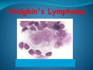
Hodgkin lymphoma • cell of origin: germinal centre B-cell • Reed-Sternberg cells (or RS variants) in the affected tissues • most cells in affected lymph node are polyclonal reactive lymphoid cells, not neoplastic cells
memory B-cell CLL MCL stem cell mature naive B-cell germinal center B-cell MZL CLL lymphoid precursor progenitor-B MM LBL, ALL pre-B DLBCL, FL, BL, HL immature B-cell plasma cell Bone marrow Lymphoid tissue B-cell development
A possible model of pathogenesis loss of apoptosis transforming event(s) EBV? cytokines germinal centre B cell RS cell inflammatory response
The Scream, 1893 Edvard Munch Reed-Sternberg cell
RS cell and variants classic RS cell lacunar cell popcorn cell (lymphocyte predominance) (mixed cellularity) (nodular sclerosis)
Hodgkins Lymphoma: • Painless, firm lymphadenopathy, • Fever* Eosinophilia • Only Reed-Sternberg cells malignant (B cell) • Classification(WHO): Classic Hodgkins: • Lymphocyte predominant. • Nodular Sclerosis. • Mixed cellularity. • Lymphocyte depleted. • Nodular lymph. predominant (non-classic)
Nodular Sclerosing < 80% Supraclavicular & mediastinal Stage I&II b From, Diagnostic Surgical Pathology of the Head and Neck, W.B.Saunders, p 750 & 764. Lymphocyte Predominant 5 % Cervical LN Stage I &II a Hodgkin’s Disease
Mixed Cellularity > 20 % Retroperitoneal Stage II & III From, Diagnostic Surgical Pathology of the Head and Neck, W.B.Saunders, p 750. Lymphocyte Depleted < 5 % Extra nodal system Stage III & IV Hodgkin’s Disease
Hodgkin’s Disease • Presentation • Asymmetric lymphadenopathy—90% • Firm, rubbery • Supraclavicular fossa • Spleen, liver (extranodal sites relatively uncommon except in advanced disease • Constitutional symptoms—1/3 of cases • Fever, night sweats, anorexia, weakness, weight loss
Lymphadenopathy in HL • Number one or two groups • Site mostly cervical • Size usually small • Shape discrete • Consistency india rubbery or firm • Mobile • No skin involvement • No tenderness • No fixation
Lymphadenopathy in NHL • Number multiple • Site mostly extra nodal • Size usually large • Shape matted • Consistency hard & cystic • Fixed • skin stretched & red • tender • fixation
HL Number one or two groups Site mostly cervical Size usually small Shape discrete Consistency india rubbery or firm Mobile No skin involvement No tenderness No fixation NHL Number multiple Site mostly extra nodal Size usually large Shape matted Consistency hard & cystic Fixed skin stretched & red tender fixation Lymphadenopathy in Lymphoma
Extranodal manifestations • SVC compression --- • dilated Neck veins (1) Cervical lymphadenopathy (b) RLN ---hoarsness of voice (3) Mediastinal (2) Hilar LN bronchial compression which cause segmental atelectasis (c) Trachea & bronchi--- cough& dyspnea (d) Lung--- Dyspnea & effusion (4)Splenomegally dt infiltration or hyperplasia • (5) Hepatomegally--- Ascites dt • Hepatic dysfunction • Peritoneal invasion (7) Stomach & bowel malabsorption syndromes • (8) Bone deposites • Sever pain • Pathological fractures (6) jaundice • Prehepatic • hemolytic autoimmune • hypersplenism • (9) Neurological • cord compression • Cranial nerve palsy • Root pains (12) Anaemia Hypersplenism BM infiltration Cytotoxic drugs • Hepatic– • cholestatic • hepatitis (10) Skin nodules (11) Mycosis fungoids Posthepatic – LN at porta hepatis
Biologically rational classification Clinically useful classification • Diseases that have distinct • morphology • immunophenotype • genetic features • clinical features • Diseases that have distinct • clinical features • natural history • prognosis • treatment The challenge of lymphoma classification
Stage I Stage II Stage III Stage IV Staging of lymphoma A: absence of B symptoms B: fever, night sweats, weight loss
Lymphoma Row of enlarged lymph nodes
Hodgkin’s Disease • Evaluation • H&P • Biopsy = Reed-Sternberg cells • Staging w/u • Similar to NHL • Laparotomy • Controversial From, Principles and Practice of Pediatric Oncology, Lippincott Williams & Wilkins, P 640.
Hodgkin’s Disease • Localized disease • Extended field XRT • Disseminated disease • MOPP = nitrogen mustard, vinblastine, procarbazine, prednisone • ABVD = adriamycin bleomycin, vincristine, dacarbazine
Laboratory Diagnosis: • Haematological: • Normocytic normochromic anemia, High ESR* • Leucocytosis, Eosinophilia, lymphopenia • Leukoerythroblastic picture - BM infiltration* • Bonemarrow: • Normal, or late involvement. • Trephine biopsy- diffuse or follicular infiltration • Biochemical: • High serum LDH – poor prognosis • Hypercalcemia, Alkaline phosphatase, Uric acid. • Serum transaminases & Bilirubin – Liver
Laboratory Diagnosis: • Haematological: • Normocytic normochromic anemia, High ESR* • Leucocytosis, Eosinophilia, lymphopenia • Leukoerythroblastic picture - BM infiltration* • Bonemarrow: • Normal, or late involvement. • Trephine biopsy- diffuse or follicular infiltration • Biochemical: • High serum LDH – poor prognosis • Hypercalcemia, Alkaline phosphatase, Uric acid. • Serum transaminases & Bilirubin – Liver
Laboratory Diagnosis: • Immunological: • Monoclonal gammopathy –B cell NHL, Myeloma • Low normal gammaglobulins • Autoimmune hemolytic anemia – auto ab. • Karyotypic/Genetic: • t(14;18) – B cell follicular (14* heavy chain) • t(11;14) – diffuse NHL
Radiological • Chest x ray • Bone scan • Bone x ray if +ve bone scan or bone pains • CT scan of chest & abdomen & pelvis • Ga 67 scan • SPRCT • PET to evaluate residuals
LN biopsy • Must whole LN as • destruction of the architecture is of diagnostic value and • also Reed Sternberg in HL id diagnostic
Additional work up in NHL • Flow cytometry • Peripheral blood • Bone marrow detect haematological involvement • Diagnostic spinal tab in • Lymphoblastic lymphoma • Burkitt’s lymphoma • Upper GIT& small bowel series & endoscopy in S&S of GIT
Diagnostic laparotomy • Indicated only in HL stage I&IIa ( as supraclavicular enlargment = 40% abdominal involvement) • Technique • Systemic LN examination • Biopsy from suspicious LN • Splenectomy • Wedge biopsy from liver • Ovariopexy • Appendectomy • Putting silver clips at the site of involved LN
Localized disease (Stage I & II) Extended field XRT Above diaphragm -------- Mantle below diaphragm --------Inverted Y Recently IFRT + new modality chemotherapy ABVD Stage III a Extended field RT IFRT + ABVD Multi agent chemotherapy ABVD or MOPP Disseminated disease (Stage III b & IV ) MOPP = nitrogen mustard, vinblastine, procarbazine, prednisone ABVD = adriamycin bleomycin, vincristine, dacarbazine Hodgkin’s Disease