Download
1 / 36
370 likes | 725 Views
Lymphoma. D S O’Briain February 2009. Lymphadenopathy. Enlarged lymph nodes Benign: soft, mobile, tender, Malign: hard, fixed, 1. Reactive hyperplasia (lymphadenitis), benign, common 2. Metastatic malignant tumour; carcinoma, melanoma, rarely sarcoma 3. Lymphoma; Hodgkin, non-Hodgkin

E N D
Lymphoma D S O’Briain February 2009
Lymphadenopathy Enlarged lymph nodes • Benign: soft, mobile, tender, • Malign: hard, fixed, 1. Reactive hyperplasia (lymphadenitis), benign, common 2. Metastatic malignant tumour; carcinoma, melanoma, rarely sarcoma 3. Lymphoma; Hodgkin, non-Hodgkin • Extranodal lymphoid tissue may be involved by non-Hodgkin lymphoma
Malignant Lymphoma • Hodgkin Lymphoma: nodal • Non Hodgkin: nodal or extranodal • B cell: diffuse large cell • Follicular • Others • T cell (more rare) • Mycosis fungoides • Others
Lymphoma/leukaemia • Incidence: Ireland 1994-8; percentage of all cancers • Lymphoid Neoplasms, 4% of all cancers
Lymphoma vs Leukaemia • Lymphoma: grossly visible mass (in node or extranodal) • Leukaemia: no gross tumour; abnormal cells identified by microscope
Lymph Lympha (Latin): clear spring water • Lymph; clear (yellow or milky) fluid in vessels (lymphatics) which lead to nodes (lymph nodes). The lymph contains cells (lymphocytes) • Lymph is also applied to clear fluid, sap or exudate • Node (Latin nodus) a knot, knob or lump • Related words: nodule, nodose—containing nodes or lumps • Follicles (Latin follis; a bellows or sac) little sacs, nodular Contrast with diffuse; evenly spread
Myeloid Myelo (Latin): marrow, pith, medulla of: 1) spinal cord or 2) bone • 1) cord related: eg. meningomyelitis, poliomyelitis • 2) marrow related • Inflammation: osteomyelitis • Tumour: myeloma (a tumour composed of plasma cells, usually multiple tumours (multiple myeloma) or single tumour (plasmacytoma) • Cells: myeloid cells, commonest cells in marrow, include neutrophils, basophils and eosinophils
B cell neoplasms • Immature • lymphoblasts—acute lymphoblastic leukaemia • Mature cells • lymphocytes—chronic lymphocytic leukaemia • plasma cells— multiple myeloma • Large cells— large cell lymphoma • Follicles —Follicular center cell lymphoma (mantle, margin cell lymphomas) • Undefined —hairy cell and Burkitt lymphoma
Lymph node architecture • 1) sinus network • Afferent lymphatics • Subcapsular, cortical, medullary sinus • Efferent lymphatic • 2) follicles (B cells) • Primary (unstimulated) • Secondary (stimulated; (reactive) germinal centre • 3) paracortex (T cells)
Antigen processing cell • Monocytes: • histiocyte, phagocyte, macrophage • epithelioid cell, giant cell, granuloma • Interdigiting reticulum cells (T cell zone) • Dendritic reticulum cells (B cell zone) • Langerhans cells • Tumours rare: Langerhans cell histiocytosis
T and B cell development • Multiple genes are available for each portion (variable, joining, diversity regions) of the immunoglobulin molecule and the alpha, beta, gamma and delta regions of the T cell receptor gene • Gene rearrangement: for each cell, one gene is chosen at random to produce each segment of the molecule. The other genes in this segment are deleted. In a cell population a huge variety of immunoglobulin or T cell receptor molecules is thus created • Immunoglobulin development: a B cell is committed to one light chain—kappa chains outnumber lambda chains by 2:1. All cells begin with M heavy chain but may switch to G, A, D, or E. • Monoclonal proliferation: all cells have identical immunoglobulin gene rearrangements and an identical product (M band immunoglobulin). Monoclonality usually equates with malignancy. It may be identified by light chain restriction or by genetic techniques (PCR, Southern blotting)
The reactive lymph node • Usually a morphologically non-specific reaction to nearby inflammation or neoplasia. • 1) follicular hyperplasia: occurs in rheumatoid arthritis, secondary syphilis, persistent generalized lymphadenitis (HIV) • Variety: giant lymph node hyperplasia (Castleman disease) • 2) sinus hyperplasia: occurs as a drainage reaction (to inflammation or carcinoma) • Variety: sinus histiocytosis with massive lymphadenopathy (Rosai Dorfman disease) • 3) paracortical hyperplasia: dermatopathic (reaction to a rash) • 4) mixed pattern (1, 2, and 3 above) a frequent non-specific finding • 5) diffuse pattern: architecture obscured (usually viral infections)
Diagnostic lymph node biopsies of peripheral lymph nodes (180 cases) • Reactive lymph node 1. toxoplasmosis, infectious mononucleosis, dermatopathic lymphadenitis, necrotising lymphadenopathy, tuberculosis, sarcoidosis 2. Adjacent tumour or infection, collagen disease
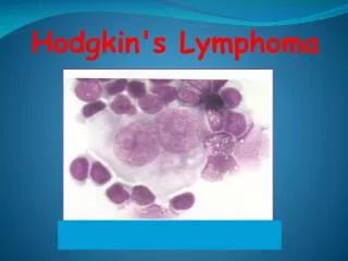
Hodgkin Lymphoma • One-third (?) of malignant lymphomas • Epidemiology; bimodal peak 20-30 years and 50+ years, predominantly young adults • Site: involves nodes, predominantly cervical • Type: • lymphocyte predominant HL • Classic HL • Morphology: usually a small number of malignant cells surrounded by abundant reactive cells attracted by high levels of cytokine production • The malignant cells are called Hodgkin cells • (varieties: Reed Sternberg, lacunar, popcorn cells) • Reactive cells: • lymphocytes, plasma cells, polys, eosinophils, histiocytes • Aetiology unknown: ? Role of Epstein Barr virus • Pathogenesis: probable B cells with defective gene rearrangement • Prognosis: 5 year survival 80% (was 20%)
Hodgkin Lymphoma Lymphocyte predominant Hodgkin Lymphoma (5%) • Nodular proliferation of scattered large B cells (popcorn cells) • Males 30-50 years • Cervical or axillary nodes • Best survival • Monoclonal B cell disease • Stains for standard B cell markers
Hodgkin lymphoma • Classic Hodgkin Lymphoma • Composed of B cells of germinal centre origin, with somatic immuoglobulin mutations, defective Ig transcription and inactivated apoptosis • Stain for CD15 and CD30 but not B cell (CD20) or T cell (CD3) markers • 4 subtypes: before modern therapy survival related to subtype. Now survival relates mainly to stage
Hodgkin Lymphoma, subtypes • Nodular sclerosis (70%+) of classic HL • 1) Reed Sternberg cells, 2) lacunar cells, 3) broad fibrotic bands • Intermediate prognosis (grade 1); grade 2 (>25% pleomorphic cells) worse prognosis • Frequently bulky (>10 cm) nodes and mediastinal involvement • Equal male and female rates • EBV less frequent than other subtypes • Mixed cellularity • 20%. Intermediate prognosis • Lymphocyte rich • 5% best prognosis • Lymphocyte depleted • Less than 5%, worst prognosis
Hodgkin Lymphoma, Staging (Ann Arbor system) Stage • 1. Single nodal region involved • 2. Two or more lymph node regions on one side of the diaphragm • 3 Nodes on both sides of diaphragm without extranodal involvement • 4 Extranodal involvement, eg bone marrow, liver, lung A or B • A - asymptomatic • B - unexplained fever (38oC), night sweats, unexplained weight loss of more than 10% body weight
Hodgkin Lymphoma, Staging (Ann Arbor system) Stage • 1. Single nodal region involved • 2. Two or more lymph node regions on one side of the diaphragm • 3 Nodes on both sides of diaphragm without extranodal involvement • 4 Extranodal involvement, eg bone marrow, liver, lung A or B • A - asymptomatic • B - unexplained fever (38oC), night sweats, unexplained weight loss of more than 10% body weight
Lymphoma classification basis • Cell morphology • Tumour architecture • Cell function • Tumour grade • Nodal or extranodal • Clinicopathological • Molecular, flow cytometry, ancillary studies
Classification of lymphoma, B-cellModified from WHO classification 2008 • Precursor B-cell lymphoid neoplasms • (B lymphoblastic leukaemia/lymphoma (9 subtypes) • Mature B-cell neoplasms (17 further subtypes) • Small lymphocytic lymphoma (CLL) • Lymphoplasmacytic lymphoma (Waldenstrom) • Plasma cell myeloma • Follicular lymphoma • Marginal zone lymphoma • Mantle cell lymphoma • Diffuse large cell lymphoma • Hairy cell lymphoma • Burkitt lymphoma
Classification of lymphoma, T-cellModified from WHO classification 2008(WHO lists 24 subtypes; selected types below) • Precursor T-cell lymphoid neoplasms • T lymphoblastic leukaemia/lymphoma • Mature T-cell and NK-cell neoplasms • Mycosis fungoides/Sezary syndrome • Peripheral T cell lymphoma NOS • Enteropathy associated T-cell lymphoma • Adult T-cell leukaemia/lymphoma • Anaplastic large cell lymphoma
Precursor Lymphoblastic lymphoma/leukaemia • B cell • 80% of acute lymphoblastic leukaemia • Frequently children • Rarely presents as tissue mass (in skin, bone, nodes) • T cell • 15% of acute lymphoblastic leukaemia • Often presents with thymic (mediastinal) mass or enlarged lymph nodes • Adolescent males
Small lymphocytic lymphoma/CLL • Most frequent leukaemia • Older adults • Marrow and peripheral blood involved • Usually some involvement of nodes, spleen and liver • WCC normal in SLL, raised in CLL • Long survival • Some markers (Zap70, CD38) predict shorter survival
Follicular lymphoma • Very frequent form of nodal lymphoma • Adults >40 years, male=female • Often widespread disease at diagnosis: nodes, marrow, spleen, sometimes also extranodal sites • Mixture of small and large cells in follicular pattern • Mostly small cells: (mean 8-10 year survival) • Mostly large cells: (mean 2-4 years survival) • t(14-18) in most (bcl-2 translocation with Ig heavy chain gene)
Diffuse large B cell lymphoma • Very frequent form of nodal and extranodal lymphoma • Large cells (centroblasts, immunoblasts, polylobated, anaplastic) • Heterogenous category (includes several different forms of lymphoma) • All ages but mainly older adults • Often single site involved (may involve multiple sites or extranodal sites) • Aggressive lymphoma • May transform from low grade lymphoma
Burkitt lymphoma • Aggressive • Vacuolated blast cells, frequent apoptotic cells, starry-sky histiocytes • Male children, jaw or abdominal mass • EB virus in Africa, but not in west • c-myc (chr 8) translocation: t(8-14)
Hairy cell leukaemia • Peculiar B cells, with hairy cytoplasmic processes • Cells have tartrate resistant acid phosphatase (by cytochemistry) and ribosome lamellar complexes (by EM) • Spleen based, involves marrow • Presents with pancytopenia and splenomegaly • Adults • Long survival, responds well to forms of chemotherapy
Plasmacytic and plasmacytoid lymphomas • Plasma cell (multiple) myeloma • Marrow plasmacytosis • Lytic or osteopenic bone lesions • Monoclonal gammopathy, other immunoglobin reduced • Urine light chains (Bence Jones) • Amyloid • Renal disease (hypercalcaemia, protein casts, amyloid • Related lesions and gammopathies • Local plasmacytoma • Monoclonal gammopathy of uncertain significance • Other lymphomas may produce M band • Waldenstrom macroglobulinaemia • Lymphoplasmacytic cells • Monoclonal gammopathy, IgM (hyperviscosity syndrome) • Hepatosplenomegaly, lymphadenopathy • Rare: immunoproliferative small intestinal disease (IgA heavy chain); IgG and IgM heavy chain diseases
Mycosis fungoides • T cell lymphoma • Older adults • Skin lymphoma: rash, plaques, nodules • Small cerebreform cells infiltrate dermis/epidermis (Pautrier pseudo or microabscess) • Leukaemic form (Sezary syndrome) • Low grade until spreads from skin as large cell lymphoma
Adult T cell leukaemia/lymphoma • HTLV-1 retrovirus • Japan or Caribbean • Generalised lymphadenopathy, hepatosplenomegaly • Skin infiltration, leukaemia, hypercalcaemia • Aggressive
Enteropathy associated T cell lymphoma • Adults (never children) • Associated with coeliac disease • Abdominal pain, obstruction, pyrexia, malabsorption • Aggressive
Extranodal lymphoma • 25-40% of non Hodgkin lymphomas arise in extranodal sites, predominantly gastrointestinal tract, and skin. Also: thyroid, salivary gland, lung, bone, orbit • Malt (mucosa associated lymphoid tissue) lymphomas. (Stomach, other GI sites, thyroid, lung, salivary gland) low grade • Enteropathy associated T cell lymphoma (coeliac, small intestine, aggressive) • Multiple lymphomatous polyposis (mantle cells, B cell, intermediate prognosis) • Immunoproliferative small intestinal disease (IgA heavy chain; intermediate prognosis) • Burkitt lymphoma; aggressive • Mycosis fungoides: slowly progressive
Lymphoma/leukaemia interface • Lymphosarcoma cell leukaemia (lymphoma cells spilling into blood) • Adult T cell leukaemia/lymphoma (HTLV, Japan, Caribbean) • Lymphoblastic lymphoma (T cell ALL with mediastinal involvement) • Richter syndrome (aggressive large cell lymphoma develops in 5% of CLL) • Sezary syndrome (circulating cerebreform T cells with skin lymphoma (mycosis fungoides) • Hairy cell leukaemia (large spleen, pancytopenia) • Granulocytic sarcoma (tumour mass of acute myeloid leukaemia cells)
Lymphomas in children • Hodgkin Lymphoma • Non Hodgkin • Burkitt • Lymphoblastic • Large cell
Molecular biology of lymphomas • Antigen receptor genes • B cell: IgH (14q32), Ig Kappa (2p12), Ig Lambda (22q11) • T cell: alpha (14q11), beta (7q34), gamma (7p15), delta (18q21) • Proto-oncogenes: bcl-1 (11q13), bcl-2 anti-apoptosis (18q21), c-myc (8q24), abl (9q34) • Translocations: are the most studied alterations in lymphoma (point mutations, gene amplifications, loss of suppressor genes are less established; but remember, tumorigenesis is a multistep process) • Most frequent translocations • t(14-18)(q32q21) IgH and bcl-2, occurs in most follicular and many diffuse lymphomas • t(8-14)(q13q32) Burkitt lymphoma • t(11-14)(q13q32) bcl-1 and IgH, mantle cell lymphoma (some CLLs) • t(9-22)(q34q11) abl and bcr, CML and some ALL • Identify malignant clone if 1-5% of cells (on southern blotting; less on PCR) are abnormal; they produce a rearranged (non-germline) band which may be confirmed by a probe