Download
1 / 105
1.12k likes | 1.67k Views
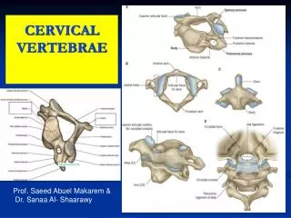
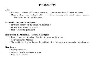
Spine (Vertebrae) Fracture And Spinal Cord Injury. Dr. Hermansyah, SpOT Bag. Bedah/ SMF Orthopedi FK-Unand/ RSUP Dr. M. Djamil Padang RSUD Lubuk Basung. Normal Spinal Anatomy. Spinal ligament. Intrasegmental Ligamentum flavum Intertransverse ligament Interspinous ligament Intersegmental

E N D
Spine (Vertebrae) Fracture And Spinal Cord Injury Dr. Hermansyah, SpOT Bag. Bedah/ SMF Orthopedi FK-Unand/ RSUP Dr. M. Djamil Padang RSUD Lubuk Basung
Spinal ligament • Intrasegmental • Ligamentum flavum • Intertransverse ligament • Interspinous ligament • Intersegmental • ALL • PLL • Supraspinous ligament
Epidemiology • Incidence: 10,000 new cases/year • Prevalence: 191,000 cases and rising • Prime occurrence: males, peak of their productive lives • Cost: $ 5.6 billion/year in the US • Cost per person: directly related to the level of SCI and patient’s age
Common Mechanisms • Compression • Flexion • Extension • Rotation • Lateral bending • Distraction • Penetration
Suspect spinal injury with... • Sudden decelerations (MVCs, falls) • Compression injuries (diving, falls onto feet/buttocks) • Significant blunt trauma (football, hockey snowboarding, jet skis) • Very violent mechanisms (explosions, cave-ins, lightning strike) • Unconscious patient • Neurological deficit • Spinal tenderness
Goal of spine trauma care • Protect further injury during evaluation and management • Identify spine injury or document absence of spine injury • Optimize conditions for maximal neurologic recovery
Goal of spine trauma care • Maintain or restore spinal alignment • Minimize loss of spinal mobility • Obtain healed & stable spine • Facilitate rehabilitation
Pre-hospital management • Protect spine at all times during the management of patients with multiple injuries • Up to 15% of spinal injuries have a second (possibly non adjacent) fracture elsewhere in the spine • Ideally, whole spine should be immobilized in neutral position on a firm surface
PROTECTION PRIORITY • Detection Secondary “Log-rolling”
Pre-hospital management • Cervical spine immobilization • Transportation of spinal cord-injured patients
Cervical spine immobilization • “Safe assumptions” • Head injury and unconscious • Multiple trauma • Fall • Severely injured worker • Unstable spinal column • Hard backboard, rigid cervical collar and lateral support (sand bag) • Neutral position
Transportation of spinal cord-injured patients • Emergency Medical Systems (EMS) • Paramedical staff • Primary trauma center • Spinal injury center
Clinical assessment • Advance Trauma Life Support (ATLS) guidelines • Primary and secondary surveys • Adequate airway and ventilation are the most important factors • Supplemental oxygenation • Early intubation is critical to limit secondary injury from hypoxia
Physical examination • Inspection and palpation • Occiput to Coccyx • Soft tissue swelling and bruising • Point of spinal tenderness • Gap or Step-off • Spasm of associated muscles • Neurological assessment • Motor, sensation and reflexes • PR • Do not forget the cranial nerve (C0-C1 injury)
Neurogenic Shock • Temporary loss of autonomic function of the cord at the level of injury • results from cervical or high thoracic injury • Presentation • Flaccid paralysis distal to injury site • Loss of autonomic function • hypotension • vasodilatation • loss of bladder and bowel control • loss of thermoregulation • warm, pink, dry below injury site • bradycardia
Neurologic assessment • Spinal shock • Bulbocavernosus reflex • Complete VS incomplete cord injury • ต้องพ้นภาวะ spinal shock ไปก่อน • Sacral sparing • Voluntary anal sphincter control • Toe flexor • Perianal sensation • Anal wink reflex
Neurologic assessment • American Spinal Injury Association grade • Grade A – E • American Spinal Injury Association score • Motor score (total = 100 points) • Key muscles : 10 muscles • Sensory score (total = 112 points) • Key sensory points : 28 dermatomes
Incomplete cord injury • Anterior cord syndrome • Brown-Sequard syndrome • Central cord syndrome
Anterior cord syndrome • Loss of motor, pain and temperature • Preserved propioception and deep touch
Brown-Sequard syndrome • Loss of ipsilateral motor and propioception • Loss of contralateral pain and temperature
Central cord syndrome • Weakness : • upper > lower • Variable sensory loss • Sacral sparing
IMAGING Numerous large prospective studies have described the large cost and low yield of the indiscriminate use of c-spine radiology in trauma patients. • WHO NEEDS AN X-RAY???
NEXUSCriteria were as follows….. • Absence of tenderness in the posterior midline • Absence of a neurological deficit • Normal level of alertness (GCS15) • No evidence of intoxication • No distracting pain elsewhere
NEXUS • Any patient who fulfilled all 5 of the aforementioned criteria were considered low risk for C-spine injury and as such did not receive C-spine radiography • For patients who had any of the 5 criteria,radiographicimaging was indicated in the form of AP, lateral, and odontoid C-spine views
Plain Film Radiology • The standard 3 view plain film series is the lateral, antero-posterior, and open-mouth view • The lateral cervical spine film must include the base of the occiput and the top of the first thoracic vertebra • The lateral view alone is inadequate and will miss up to 15% of cervical spine injuries.
X-ray Guidelines (cervical) • Adequacy, Alignment • Bone abnormality, Base of skull • Cartilage, Contours • Disc space • Soft tissue
Interpreting Lateral Plain Film • Adequacy • Should see C7-T1 junction • If not get swimmer’s view or CT
Interpreting lateral Plain Film • Alignment • Anterior vertebral line • Formed by anterior borders of vertebral bodies • Posterior vertebral line • Formed by posterior borders of vertebral bodies • Spino-laminar Line • Formed by the junction of the spinous processes and the laminae • Posterior Spinous Line • Formed by posterior aspect of the spinous processes
Cartilage • Predental Space should be no more than 3 mm in adults and 5 mm in children • Increased distance may indicate fracture of odontoid or transverse ligament injury
Cartilage Cont. • Disc Spaces • Should be uniform • Assess spaces between the spinous processes
Soft tissue • Nasopharyngeal space (C1) - 10 mm (adult) • Retropharyngeal space (C2-C4) - 5-7 mm • Retrotracheal space (C5-C7) - 14 mm (children), 22 mm (adults) • Extremely variable and nonspecific Measurements anterior to the mid-cervical spine up to 7 mm are common. > 7 mm,-a fracture is likely and the neck should be immobilized.
AP C-spine Films Spinous processes should line up Disc space should be uniform Vertebral body height should be uniform. Check for oblique fractures.
Open mouth view Adequacy: all of the dens and lateral borders of C1 & C2 Alignment: lateral masses of C1 and C2 Bone: Inspect dens for lucent fracture lines
CT Scan • Thin cut CT scan should be used to evaluate abnormal, suspicious or poorly visualized areas on plain film • The combination of plain film and directed CT scan provides a false negative rate of less than 0.1%
MRI • Ideally all patients with abnormal neurological examination should be evaluated with MRI scan
Management of SCI • Primary Goal • Prevent secondary injury • Immobilization of the spine begins in the initial assessment • Treat the spine as a long bone • Secure joint above and below • Caution with “partial” spine splinting
Management of SCI • Spinal motion restriction: immobilization devices • ABCs • Increase FiO2 • Assist ventilations as needed with c-spine control • Indications for intubation : • Acute respiratory failure • GCS <9 • Increased RR with hypoxia • PCO2 > 50 • VC < 10 mL/kg • IV Access & fluids titrated to BP ~ 90-100 mmHg