Download
1 / 70
700 likes | 923 Views
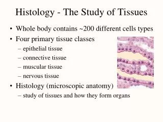
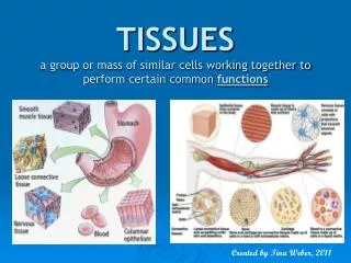
Group of similar cells common embryonic origin common function Histology study of tissues. 200 Different cell types Four primary tissue classes epithelial tissue connective tissue muscular tissue nervous tissue Histology (microscopic anatomy) study of tissues organ formation

E N D
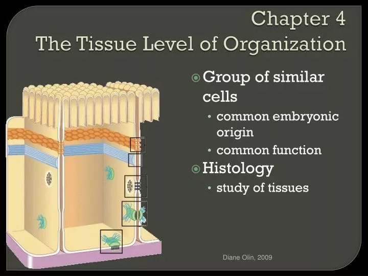
Group of similar cells • common embryonic origin • common function • Histology • study of tissues Diane Olin, 2009
200 Different cell types • Four primary tissue classes • epithelial tissue • connective tissue • muscular tissue • nervous tissue • Histology (microscopic anatomy) • study of tissues organ formation • Organ = structure with discrete boundaries • composed of 2 or more tissue types Diane Olin, 2009
Epithelial Tissue • covers surfaces because cells are in contact • lines hollow organs, cavities and ducts • forms glands when cells sink under the surface • Connective Tissue • supports and binds structures together • stores energy as fat • provides immunity to disease Diane Olin, 2009
Muscle Tissue • cells shorten in length producing movement (contracts) • Nerve Tissue • cells that conduct electrical signals • detects changes inside and outside the body • responds with nerve impulses Diane Olin, 2009
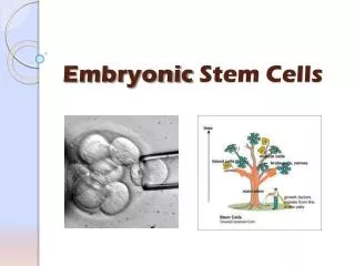
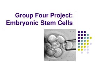
Embryo begins as single cell • divides into many cells and layers (strata) • 3 Primary germ layers • ectoderm (outer) • forms epidermis and nervous system • endoderm (inner) • forms mucous membrane lining GI tract and respiratory system and digestive glands • mesoderm (middle) becomes mesenchyme • wispy collagen fibers and fibroblasts in gel matrix • gives rise to muscle, bone, blood Diane Olin, 2009
Removal of living tissue for microscopic examination Useful for diagnosis, especially cancer Tissue preserved, sectioned and stained before microscopic viewing Diane Olin, 2009
Closely packed cells forming continuous sheets • Cells sit on basement membrane • Apical (upper) free surface • Avascular---without blood vessels • nutrients diffuse in from underlying connective tissue • Good nerve supply • Rapid cell division Diane Olin, 2009
Basal lamina • from epithelial cells • collagen fibers • Reticular lamina • secreted by connective tissue cells • reticular fibers • holds cells to connective tissue • guide for cell migration during development Diane Olin, 2009
Notice how the mitochondria are concentrated at the basal portion of cell-probably to provide energy for cell’s transport processes. Cilia are at apical portion; sweeping action needed there. By polarity we mean there is an unequal distribution of the organelles or features in the cell. It is not symmetrical either. Diane Olin, 2009
All cells with the exception of metatastic cancer cells and blood have these • They keep cells anchored to each other and to the matrix so that there can be proper growth and cell division. • They enable cells to resist stress and communicate with each other. Diane Olin, 2009
The lipid portions of two cell membranes are tightly bound together • A continuous adhesion belt forms • This connection prevents the passage of all solutes and even water between two cells. (really tight!!) Diane Olin, 2009
Blood Brain Barrier Diane Olin, 2009
Blood Brain Barrier Diane Olin, 2009
Remember these resist mechanical stress: • Differences between the desmosomes and hemidesmosomes (belt) below • Desmosomes –attach cells to each other (are “whole” bottons. • Hemidesmosomes- hemi (means half) and attaches to the basal lamina instead. This one anchors the cell in place. • Those blue strands are like cross-braces (fibers from the cytoplasm) that help in this stablilization process. Diane Olin, 2009
These are junctions of communication • They allow for the passage of molecules • The junctions are called GAP junctions • The proteins that hold the junctions together are called CONNEXONS Diane Olin, 2009
Ring of transmembrane proteins form a water-filled channel • small solutes pass directly from cell to cell • in embryos, cardiac and smooth muscle Diane Olin, 2009
Each gap junction is composed of a 6-sectioned monomer protein “connexon”. Notice the open and closed configurations on the right. Diane Olin, 2009
Moves fluids over epithelial tissue • Moves fluids through epithelial tissue OR • Produces secretions that provide physical protection or act as chemical messengers. Diane Olin, 2009
Covering and lining epithelium • epidermis of skin • lining of blood vessels and ducts • lining respiratory, reproductive, urinary & GI tract • Glandular epithelium • secreting portion of glands • thyroid, adrenal, and sweat glands Diane Olin, 2009
Classified by arrangement of cells into layers • simple = one cell layer thick • stratified = many cell layers thick • pseudostratified = single layer of cells where all cells don’t reach apical surface • nuclei at found at different levels so it looks multilayered • Classified by shape of surface cells • squamous =flat • cuboidal = cube-shaped • columnar = tall column • transitional = shape varies with tissue stretching Diane Olin, 2009
Single layer of flat cells • lines blood vessels (endothelium), body cavities (mesothelium) • very thin --- controls diffusion, osmosis and filtration • nuclei centrally located • Cells in direct contact with each other Diane Olin, 2009
Surface view of lining of peritoneal cavity • Section of intestinal showing serosa Diane Olin, 2009
Single layer of cube-shaped cells viewed from the side • Nuclei round and centrally located • Lines tubes of kidney • Absorption or secretion Diane Olin, 2009
Sectional view of kidney tubules Diane Olin, 2009
Single layer rectangular cells • Unicellular glands =goblet cells secrete mucus • lubricate GI, respiratory, reproductive and urinary systems • Microvilli = fingerlike cytoplasmic projections • for absorption in GI tract (stomach to anus) Diane Olin, 2009
Section from small intestine Diane Olin, 2009
Single layer rectangular cells with cilia • Mucus from goblet cells moved along by cilia • found in respiratory system and uterine tubes Diane Olin, 2009
Section of uterine tube Diane Olin, 2009
Several cell layers thick • Surface cells flat • Keratinized = surface cells dead and filled with keratin • skin (epidermis) • Nonkeratinized = no keratin in moist living cells at surface • mouth, vagina Diane Olin, 2009
Section of vagina Diane Olin, 2009
Multilayered • Surface cells cuboidal • rare (only found in sweat gland ducts & male urethra) Diane Olin, 2009
Multilayered • Surface cells columnar • Rare (very large ducts & part of male urethra) Diane Olin, 2009
Multilayered Surface cells varying in shape from round to flat if stretched Lines hollow organs that expand from within (urinary bladder) Diane Olin, 2009
Single cell layer All cells attach to basement membrane but not all reach free surface Nuclei at varying depths Respiratory system, male urethra & epididymis Diane Olin, 2009
Derived from epithelial cells that sank below the surface during development Diane Olin, 2009
EXOCRINE and ENDOCRINE • Exocrine: have a duct and release their secretions through the duct onto an epithelial surface. • Endocrine : Their secretions are released directly into the interstitial fluid. Usually these are hormones. We will not consider these in this course. But rather, focus on exocrine glands. Diane Olin, 2009
Pancreas contains both EXOCRINE and ENDOCRINE cells. Beta cells (endocrine cells) secrete insulin. Acinar cells (exocrine cells) secrete Diane Olin, 2009
The duct portion may be branched (called compound) or unbranched (called simple). The glandular portion may be tubular, acinar, or may be a mix of the two (called tubuloacinar). If the glandular portion branches, then the gland is called a branched gland.
SIMPLE: Just a tube can be multicellular ; can be unicellular as in a goblet cell, COMPOUND: Branching Acinar; Means a secretory sac or aveolus Tubular: No secretory sac or aveolus NOW…put it together and review the previous slide…. Diane Olin, 2009