Download
1 / 11
120 likes | 275 Views
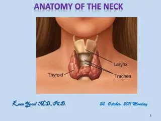
Anatomy of the thryroid. Where is it?. In the neck Inferior to larynx Anterolateral structure. 4 parathyroid glands. Branches of the aorta. A orta B rachiocephalic Left C ommon carotid Left S ubclavian artery. Dual Blood Supply. Aorta (RHS brachiocephalic Lhs direct)

E N D
Where is it? • In the neck • Inferior to larynx • Anterolateral structure
Branches of the aorta • Aorta • Brachiocephalic • Left Common carotid • Left Subclavian artery
Dual Blood Supply • Aorta • (RHS brachiocephalic • Lhs direct) • common carotid • external carotid • superior thyroid artery • (RHS brachiocephalic, LHS aorta) • Subclavian • thyrocervical trunk • inferior thyroid artery
Venous drainage • Superior & middle thyroid drain to internal jugular (to subclavian to brachiocephalic) • Inferior thyroid into brachiocephalic veins
Embryology • Starts life on the tongue and descends via the thyroglossal duct Thyroglossal duct cysts Can form anywhere along the thyroglossal duct in the midline neck. Move on swallowing and tongue protrusion The thyroid gland and lumps tends to move during swallowing but not always with tongue protrusion.
T4 TSH Iodine deficiency • high TSH • low T4 + decreased T4 secretion Increased gland growth Adapted from Endocrinology Physiology. SP Porterfield, Mosby
T4 TSH Graves disease • Low TSH • high T4 - - + Increased T4 secretion TSI thyroid stimulating immunoglobulin Increased gland growth Adapted from Endocrinology Physiology. SP Porterfield, Mosby