Download
1 / 21
230 likes | 457 Views
Biology of the T lymphocyte. Nature of T cell - overview. T cells have a receptor for Ag on surface (TCR) which share similarities for Ig receptor on B cells; but differs: Ig can interact with Ag directly (even in solution), yet TCR recognize Ag when bound to MHC molecules.

E N D
Nature of T cell - overview • T cells have a receptor for Ag on surface (TCR) which share similarities for Ig receptor on B cells; but differs: Ig can interact with Ag directly (even in solution), yet TCR recognize Ag when bound to MHC molecules. • End product of B cell recognition = Ab; T cell recognition • 1) cytokine production • 2) destruction of cells with foreign Ag • Both T & B cells may be activated, proliferate & differentiate to memory cells
T cell populations must be able to respond to many foreign antigens, yet NOT bind to self Ag;At least four mechanisms involved; • Basic TCR repertoire determined by number of different germline genes • T cells whose TCR bind to self Ag destroyed within thymus • Surviving T cell populations expanded by positive selection in thymus followed by interaction between TCR and MHC molecules • Peripheral T cell numbers increased by exposure to foreign Ag
T cells ab gd CD4+ CD8+ Helper 1 Helper 2 Cytotoxic Regulatory Antigen receptors Accessory molecules Functions * T cell expresses ab+gd- or ab-gd+ but not ab+gd+.
CDR1, 2 & 3 NOT T cell specific Figure 9.1 The structure of the T-cell receptor (TCR) complex showing the predominant form of the antigen-binding chains, a and b, and the associated signal transduction complex, CD3 (g, d, and e chains) plus z or h. ITAMs are indicated by the rectangular boxes.
CD4 & CD8 • Coreceptor or accessory molecules • T cells: 50 - 60% CD4+, 20 - 25% CD8+ • Two functions • Adhesion molecules : Bind to MHC (CD4 binds to MHC II, CD8 binds to MHC I) --- tighten binding of T cells to APC • Signal transducers : CD4 & CD8 phosphorylated when Ag bound to ab TCR
Figure 9.2 The TCR coreceptors and their interaction with MHC molecules. (A) CD4 and (B) CD8.
Figure 9.3 The interaction of TCR, MHC, and peptide. The complementarity determining regions (CDRs) of the TCR V regions and peptide bound in the peptide-binding groove of an MHC class I molecule are depicted. [Based on the crystal structure described by K. C. Garcia et al. (1998): Science 279: 1166.]
Figure 9.4 Organization of the a, b, and d genes coding for the human T-cell receptor. The organization of the g-gene locus is not shown because of its complexity.
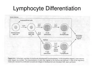
Figure 9.5 The developmental pathways of T cells in the thymus. Genes coding for the a, b, g, and d chains of the T-cell receptor are designated as a0 etc. if unrearranged and a+ if rearranged.
Figure 9.6 Positive and negative selection of ab+CD4+CD8+ T cells in the thymus.