Download
1 / 59
1.05k likes | 2.48k Views
Chest Tubes. by Charlotte Cooper RN, MSN, CNS modified by Kelle Howard RN, MSN Modified by Darlene “Cookie” Wilson, RN, MSN. Thoracic Cavity. Lungs Mediastinum Heart Aorta and great vessels Esophagus Trachea. Breathing: Inspiration. Diaphragm contracts Moves down

E N D
Chest Tubes by Charlotte Cooper RN, MSN, CNS modified by Kelle Howard RN, MSN Modified by Darlene “Cookie” Wilson, RN, MSN
Thoracic Cavity • Lungs • Mediastinum • Heart • Aorta and great vessels • Esophagus • Trachea
Breathing: Inspiration • Diaphragm contracts • Moves down • Increasing the volume of the thoracic cavity • When the volume increases, the pressure inside ________. • aka? • Pressure within the lungs is called intrapulmonary pressure
Breathing: Exhalation • Phrenic nerve stimulus stops • Diaphragm relaxes • This ______ the volume of the thoracic cavity • Lung volume decreases, intrapulmonary pressure _____
Physics of Gases If two areas of different pressure communicate, gas will move from the area of higher pressure to the area of lower pressure
Pleural Anatomy • Parietal pleura • lines the chest wall • Visceral pleura (pulmonary) • covers the lung
Pleural Anatomy Visceral pleura Parietal pleura Lung Intercostal muscles Ribs Normal Pleural Fluid Quantity: Approx. 10-20 mL per lung
Pleural Physiology • The area between the pleura is called the pleural space (sometimes referred to as “potential space”) • Normally, vacuum (negative pressure) in the pleural space keeps the two pleura together and allows the lung to expand and contract • It is also what holds the 2 pleura’s together.
Review Questions A&P of chest (You tube http://www.youtube.com/watch?v=wKhldyCC6wg)
What is this? archive.student.bmj.com/.../02/education/52.php
Pleural Injury: Therapeutic Interventions • Diagnostic tests • Client position • Treatment depends on severity • Chest tube • Heimlich valve on chest tube
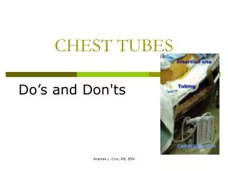
Chest Tubes • Also called “thoracic catheters” • Different sizes • From infants to adults • Small for air, larger for fluid • Different configurations • Curved or straight • Types of plastic • PVC • Silicone • Coated/Non-Coated • Heparin • Decrease friction
Chest Tube Placement • In what setting/environment is a chest tube placed? • A. Operating Room • B. Bedside • C. Emergency Room • D. All of the above • E. None of the above
Chest Tube Placement Procedure • Sterile technique • Small incision • Tube is sutured • Dressing applied • What type? Can the client have 2 chest tubes? When the tube is placed, is it clamped or unclamped? why or why not?
Chest tube insertion Choose site Suture tube to chest Explore with finger Place tube with clamp Photos courtesy trauma.org\
Heimlich ValveWhat are some problems with this valve? http://www.scielo.br/img/revistas/jbpneu/v34n8/en_a04fig01.gif
Prevent air & fluid from returning to the pleural space Chest tube is attached to a drainage device • Allows air and fluid to leave the chest • Contains a one-way valve to prevent air & fluid returning to the chest • Designed so that the device is below the level of the chest tube for gravity drainage
Treatment goal for pleural injuries 1. Remove fluid & air as promptly as possible 2. Prevent drained air & fluid from returning to the pleural space 3. Restore negative pressure in the pleural space to re-expand the lung
Interventions • Dressing changes • When? • No dependent loops • What is this? • Why? • Oxygen therapy • Record output • How often? • Analgesics • ***IS and turn, cough, deep breathe
Nursing assessment and pertinent nursing problems/interventions • Health history-respiratory disease, injury, smoking, progression of symptoms • Physical exam- degree of apparent resp distress, lung sounds, O2 sat, VS, LOC, neck vein distention, position of trachea • All require observation for respiratory symptoms • Pertinent nursing problems • Acute pain • Ineffective airway clearance • Impaired gas exchange • Home care
How a chest drainage system works
Purpose of Chamber One: • Correct any fluid comingback from the patient. • Purpose of Chamber Two: • Water seal, one-way valveallows air out of the patientbut not back into the patient. • Purpose of Chamber Three • Suction control. Manual or Fluid filled (most hospital systems)
Prevent Air and Fluid Backflow Tube open to atmosphere vents air Tube from patient
Restore negative pressure in the pleural space The depth of the water in the suction bottle determines the amount of negative pressure that can be transmitted to the chest, NOT the reading on the vacuum regulator
How a chest drainage system works • Expiratory positive pressure • Gravity • Suction
How a chest drainage system works • Expiratory positive pressure from the patient helps push air and fluid out of the chest (cough, Valsalva) • Gravityhelps fluid drainage as long as the chest drainage system is below the level of the chest • Suction can improve the speed at which air and fluid are pulled from the chest
From bottles to a box From patient To suction from patient Suction control bottle Water seal bottle Collection bottle Suction control chamber Water seal chamber Collection chamber
Water suction on left Dry suction on rightLewis p. 589 Fig 28-8
Atrium Chest Tube System • Chamber A • Suction control chamber • How do you know what level the water should be at? • Chamber B • Water seal chamber • How do you know what level the water should be at? • Should the ball be fluctuating in this chamber? • What if it isn’t? • Chamber C • Air leak monitor • What does bubbling mean? • Chamber D • Collection chamber • When do you record output? Be sure you under stand how to set up the system, the function of each chamber and how to troubleshoot issues with each chamber.
Monitoring • Water seal is a window into the pleural space • Not only for pressure • If air is leaving the chest through an air leak, bubbling will be seen here • Air meter (1-5) provides a way to “measure” the air leaving and monitor over time – getting better or worse?
Assessment • Focused respiratory assessment • Breath sounds • Respiratory rate • Respiratory depth • SpO2 • ABG • CXR
Assessment • Cardiovascular assessment • Level of consciousness • Pain • Chest tube • Amount of drainage • Insertion site & dressing
Interventions r/t chest tubes • System position • Tubing position • What happens when the patient lays on it? • Connections to patient and system • Assessing the system • Monitoring output
Complications What are some common complications?
Complications & Troubleshooting • Chest tube malposition (most common) • Subcutaneous emphysema • What is this? • What are some nursing interventions related to this complication? • High Fluid in Water Seal Chamber • Chest system may need to be vented • Air leak • How do you know? • What do you do? Others pleural effusion, inc. pneumo mediastinal shift Do chest tubes get clotted off? What can happen when fluid is removed too fast?