Download
1 / 136
1.4k likes | 1.89k Views
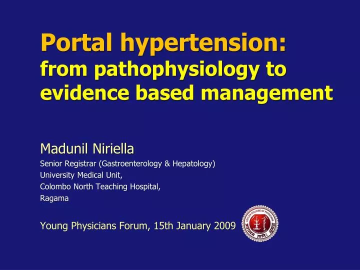
Portal hypertension: from pathophysiology to evidence based management. Madunil Niriella Senior Registrar (Gastroenterology & Hepatology) University Medical Unit, Colombo North Teaching Hospital, Ragama Young Physicians Forum, 15th January 2009 . Case History 1.

E N D
Portal hypertension: from pathophysiology to evidence based management Madunil Niriella Senior Registrar (Gastroenterology & Hepatology) University Medical Unit, Colombo North Teaching Hospital, Ragama Young Physicians Forum, 15th January 2009
Case History 1 • 37 year-old male • Chronic alcoholic liver disease – 2 years • Presented with an acute UGI bleed • Hypovolaemic shock on admission • Failed resuscitation • Expired within one hour of admission
Case History 1 cont. • History of several admissions with decompensation • No UGIE previously • Not on prophylaxis • No regular follow up • Continued to abuse alcohol
Case History 1 cont. • History of several admissions with decompensation • No UGIE previously • Not on prophylasix • No regular follow up • Continued to abuse alcohol Absence of screening and primary prophylaxis
Case History 2 • 33 year-old male • Cryptogenic cirrhosis – 8 months • Primary prophylaxis with EVB for diagnosed oesophageal varices one week back • Presented with first episode of acute UGI bleed
Case History 2 cont. • Hypovolaemic shock on admission • Resuscitated with I.V. fluids and blood • Underwent emergency UGIE
Case History 2 cont. • UGIE revealed active bleeding from ulcer sites from previous banding • Bleeding arrested by injection of 1:10000 adrenaline • Recovered following multiple blood transfusions with packed red cells, FFP, and i.v. infusions of omeprazole and terlipressin for 72 hours
Case History 2 cont. • UGIE revealed active bleeding from ulcer sites from previous banding • Bleeding arrested by injection of 1:10000 adrenaline • Recovered following multiple blood transfusions with packed red cells, FFP, and i.v. infusions of omeprazole and terlipressin for 72 hours Inappropriate prophylaxis ?
“Management of portal hypertension and its complications iscomplex and challenging”
Objectives • What is portal hypertension (PH) ? • What is the natural history and prognosis ? • How to diagnose PH ? • What is the rational basis for therapy ? • How to prevent the first bleeding episode ? • How to prevent recurrent bleeding episodes ?
Objectives • What is portal hypertension (PH) ? • What is the natural history and prognosis ? • How to diagnose PH ? • What is the rational basis for therapy ? • How to prevent the first bleeding episode ? • How to prevent recurrent bleeding episodes ?
The history of PH • 1543 - Andreas Vesalius First anatomical picture of the portal venous system
The history of PH • 1650s - Francis Glisson • Established the function of the portal vein • 1700s – Giovanni Batista Morgagni • Described varices in the splenic and short gastric veins • 1832 – Jean Cruveilhier Described the clinical picture of splenomegaly, ascites and gastrointestinal haemorrhage • 1841 - Philbert Constant Sappey Described porto-systemic collaterals
The history of PH • 1902 - Augustin Nicolas Gilbert Introduced the term “Portal hypertension”
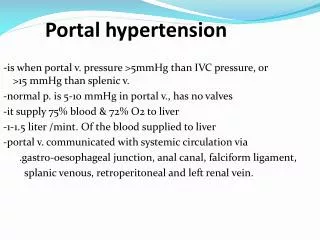
Definition of PH • Pathological increase of the portal pressure gradient (PPG) PPG = (Portal venous pressure – IVC pressure) PPG > upper normal value (5 mmHg) • Formation of portal-systemic collaterals Shunting of portal blood flow to systemic circulation bypassing the liver
HVPG: what is it? (Internal zero)
Spectrum of HVPG 3-5 mmHg 6-9 mmHg ≥10 mmHg
Clinically significant PH (CSPH) • In cirrhosis cases • Histologically proven • Well-compensated • At the time of diagnosis • 60% will have CSPH
Causes of PH • Classified according to site of obstruction to blood flow • Pre-hepatic • Portal vein thrombosis • Intra-hepatic • Cirrhosis (90%) • Pre-sinusoidal • Sinusoidal • Post-sinusoidal • Post-hepatic • Hepatic vein thrombosis (Budd-Chiari syndrome)
Causes of PH • Classified according to site of obstruction to blood flow • Pre-hepatic • Portal vein thrombosis • Intra-hepatic • Cirrhosis (90%) • Pre-sinusoidal • Sinusoidal • Post-sinusoidal • Post-hepatic • Hepatic vein thrombosis (Budd-Chiari syndrome)
Clinical manifestations • Cirrhotic PH – “Vascular disease” • Involves several systems and organs • “A Multi-organ Disease”
Multi-organ involvement • Splanchnic vascular bed – • Formation of porto-systemic collaterals • Brain – • HE • Heart – • Cirrhotic • cardiomyopathy • Systemic circulation -Hyperkinetic syndrome • Lung – • Hepato-Pulmonary • Syndrome • Kidneys - • Hepato-Renal Syndrome • Blood - • Splenomegaly and hypersplenism
Multi-organ involvement Variceal haemorrhage is the most common lethal complication of cirrhosis • Splanchnic vascular bed – • Formation of porto-systemic collaterals • Brain – • HE • Heart – • Cirrhotic • cardiomyopathy • Systemic circulation -Hyperkinetic syndrome • Lung – • Hepato-Pulmonary • Syndrome • Kidneys - • Hepato-Renal Syndrome • Blood - • Splenomegaly and hypersplenism
Multi-organ involvement Variceal haemorrhage is the most common lethal complication of cirrhosis • Splanchnic vascular bed – • Formation of porto-systemic collaterals • Brain – • HE • Heart – • Cirrhotic • cardiomyopathy • Systemic circulation -Hyperkinetic syndrome • Lung – • Hepato-Pulmonary • Syndrome Varices and variceal haemorrhage are the complications of cirrhosis that result most directly from PH • Kidneys - • Hepato-Renal Syndrome • Blood - • Splenomegaly and hypersplenism
Porto-systemic collaterals • Decompress the portal circulation • by shunting blood to systemic circulation • Sites Distal oesophagus and proximal stomach • Gastroesophageal varices (GOV) • Major collaterals – largest flow via short and left gastric veins Rectum • Rectal varices Umbilicus • Caput medusa Retroperitoneum
Objectives • What is portal hypertension (PH) ? • What is the natural history and prognosis ? • How to diagnose PH ? • What is the rational basis for therapy ? • How to prevent the first bleeding episode ? • How to prevent recurrent bleeding episodes ?
Natural History • Progressive increase in HVPG • Chain of events • Development of varices • Progressive dilatation of varices • Rupture and bleeding of varices
Pathogenesis of variceal bleeding ........
Prevalence and formation of varices • At the time of diagnosis • Compensated cirrhosis • 30-40% have varices • Decompensated cirrhosis • 60% have varices • Annual incidence of new varices (de novo) • 7% (5-10%) per annum • Appearance • HVPG > 10 mmHg • Strong predictor for development of varices
Prevalence of varices in patients with cirrhosis without prior bleeding
Progression of varices from small to large • Rate or progression • 5 to 30% per year • Predictors of progression • Child-Pugh class (most consistent) • Increase in HVPG • Alcoholic aetiology • Presence of red wale marks
Incidence of first bleeding • Overall • Incidence 4% per year • Large varices • Incidence 15% per year
Risk indicators of first bleeding • Rupture and bleeding • Large varices • HVPG > 12 mmHg • Child-Pugh class C • Presence of red-signs • Red wale marks (longitudinal red streaks on varices) • Cherry-red spots (red discrete flat spots on varices) • Haematocystic spots (discrete, red raised spots) • Diffuse erythaema
Prognosis after a bleed • 1/3 cirrhotic experience variceal bleeding • Each episode • 15-20% mortality at 6 weeks • Untreated • Rebleeding occurs in 60% within 1-2 years • 70% die < one year on the initial bleed
Objectives • What is portal hypertension (PH) ? • What is the natural history and prognosis ? • How to diagnose PH ? • What is the rational basis for therapy ? • How to prevent the first bleeding episode ? • How to prevent recurrent bleeding episodes ?
Diagnostic modalities • Invasive • HVPG measurement • Endoscopy • Endoscopic video capsule • Non-invasive • Clinical signs and lab findings • Imaging techniques • Liver stiffness (Fibroscan)
HVPG • Invasive • Measured by hepatic vein catheterization • Gold standard • Objective and quantitative equivalent of PPG in cirrhosis • Add prognostic information • Compensated cirrhosis • Acute variceal bleeding • Liver transplantation
HVPG - limitations • Lack of local expertise • Poor adherence to guidelines • Cannot ensure reliable and reproducible measurements • Invasive • Cost
Endoscopy • At time of diagnosis of cirrhosis • All patients • To document the presence of varices • To determine the risk for variceal haemorrhage • To detect patients requiring prophylactic treatment
Endoscopy screening for GOV • Without varices • Small varices • Child class A • No Red signs Rescreen every 2-3 years At the time of hepatic decompensation
Endoscopy screening for GOV • Small varices • Child class B or C • Red wale marks • Evidence of hepatic decompensation Repeat screening at 1-year intervals
Endoscopic video-capsule • Repeated conventional endoscopies • Intolerant • Capsule endoscopy • Improve patient tolerance • Once swallowed records images at pre determined intervals
Endoscopic video-capsule • Allows correct identification of varices in 80% cases • Problems • Not good at assessing variceal size • Poor accuracy in identifying • Gastric varices • Portal hypertensive gastropathy • Not recommended as the routine screening method for GOV
Non-invasive tests • Ideal test to diagnose and follow-up PH • Reproducible • Inexpensive • Non-invasive • No non-invasive procedure proved to be accurate enough to avoid endoscopy in patients with negative indicators