Download
1 / 70
700 likes | 888 Views
Muscular System Chapter 6. MUSCULAR SYSTEM. Total muscles in human body: 650 Muscles required to smile: 17 Muscles required to frown: 33 Muscles required to walk: at least 200 Longest muscle: Gluteus maximus ( butt muscle) Shortest muscle: ear muscle. Characteristics of Muscles.

E N D
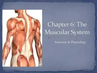
Muscular System Chapter 6
MUSCULAR SYSTEM • Total muscles in human body: 650 • Muscles required to smile: 17 • Muscles required to frown: 33 • Muscles required to walk: at least 200 • Longest muscle: Gluteus maximus ( butt muscle) • Shortest muscle: ear muscle
Characteristics of Muscles • Muscle cells are elongated (muscle cell = muscle fiber) • Contraction of muscles is due to the movement of microfilaments (actin and myosin) • All muscles share some terminology • Prefix myo refers to muscle • Prefix mys refers to muscle • Prefix sarco refers to flesh
Functions of Muscular system • Contract: • Skeletal Muscles: body movement • Smooth muscles: movement of contents through blood vessels, digestive organs • Cardiac Muscles: Pumping of heart • Generate heat : generate heat, by product of ATP needed for muscle contraction • Stabilize joints: Skeleton muscles pull on bones for movement, also stabilize joints • Maintaining Posture: Keeps the body straight against gravity
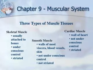
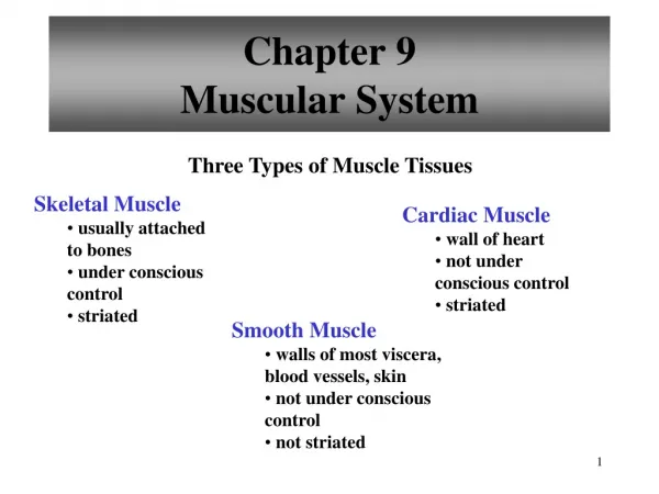
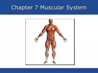
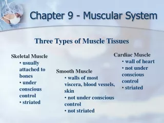
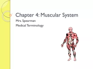
The Muscular System • Muscles are responsible for all types of body movement • Three basic muscle types are found in the body • Skeletal muscle • Cardiac muscle • Smooth muscle
Smooth Muscle • Smooth muscles are non striated, uni-nucleated and spindle shaped • Involuntary • Found in the walls of visceral organs, eg. Stomach, urinary bladder, respiratory passages
Smooth Muscles • Arranged in two layers: • circular layer • longitudinal layer • These two layers alternately contract and relax • And move food through digestive tract, emptying the bowels & bladder • Maintain housekeeping activities • Slow and steady
Cardiac Muscle • Responsible for heart pumping • Cardiac muscles are striated, cylindrical, uninucleated & branched • The muscle is under involuntary control and contract at regular intervals
Cardiac Muscle • Cardiac muscle cells are joined to another cell by intercalated discs • Discs are important in transport of impulses • Present in heart only • Cardiac fibers are arranged in spiral or eight-shaped bundles
Skeletal Muscle • Under voluntary or CNS control • Respond to impulses • Muscles are striated (actin and myosin are alternatively arranged) • Muscle cells are cylindrical or cigar shaped • multinucleated
Skeletal Muscle • Attached to bones with the help of tendons • A person has same number of muscle fiber from his birth to adult hood • However, their size increases or decreases depending on the usage
Structure of Skeletal Muscle • Each muscle fiber is enclosed in a connective tissue called endomysium • Each muscle fiber is supplied with capillary and nerve cell extensions • Several muscle fibers (cells) called fascicle are enclosed in a connective tissue called perimysium
Structure of Skeletal Muscle • Several fascicles are surrounded by third or heavy layer of connective tissue layer called epimysium • Thus each skeletal muscle is surrounded by epimysium • In order to form a long muscle, muscle fibers are arranged end to end
Skeletal Muscle Attachments • Epimysium blends into a connective tissue attachment • Tendons — cord-like structures • Mostly collagen fibers • Aponeuroses — sheet-like structures • Attach muscles indirectly to bones, cartilages, or connective tissue coverings
Microscopic Anatomy of Skeletal Muscle • Skeletal cells are multinucleate • Outer boundary of the cell is made of plasma membrane – sarcolemma • Cytoplasm of muscle cell - sarcoplasm • Endoplasmic reticulum – Sarcoplasmic reticulum • Sarcoplasm is packed with myofibrils • Other organelles, such as mitochondria, glycogen granules are found between myofibrils
Microscopic Anatomy of Skeletal Muscle • Myofibrils: are thread like organelles • Composed of protein threads called myofilaments: • thin (actin) • thick (myosin) • Sarcomeres: repeating units of myofilaments • Interaction between actin and myosin filaments leads muscle to shorten or contract • The tropomyosin/troponin complex regulates the interaction between actin and myosin
Structure of Sarcomere • Repeating units of myofibrils - Sarcomere • The distance between two Z discs is the sarcomere • The point where actin originates is called Z disk • Each sarcomere has alternating actin and myosin filaments
Structure of Sarcomere • The arrangement of myosin (dark in color and is called anisotropic band or A band) • And actin (light in color and is called isotropic or I band) alternatively • gives the muscle a striated appearance • H zone (bare zone) - lacks actin filament
Microscopic Anatomy of Skeletal Muscle • Myosin filaments have heads (extensions, or cross bridges) • When contraction occurs, actin and myosin overlap
Properties of Skeletal Muscle Activity • Irritability – ability to receive and respond to a stimulus • Contractility – ability to shorten when an adequate stimulus is received
Nerve Stimulus and Action Potential • Skeletal muscles must be stimulated by a nerve to contract • One motor neuron can stimulate many muscle cells – Motor unit • Axon of the neuron branches into many axon terminals at Neuromuscular junctions • Neuromuscular junctions – association site of nerve and muscle
Nerve Stimulus and Action Potential • Synaptic cleft – gap between nerve and muscle • Nerve and muscle do not make contact • Area between nerve and muscle is filled with interstitial fluid
Nerve Stimulus and Action Potential • When nerve impulse reaches the axon terminal, it releases the chemical known as Neurotransmitter • The neurotransmitter for skeletal muscle is acetylcholine • Neurotransmitter attaches to receptors on the sarcolemma • Sarcolemma becomes permeable to sodium (Na+)
Nerve Stimulus and Action Potential • Sodium rushing into the cell generates an action potential • Once started, muscle contraction cannot be stopped • Stimulates release of Ca+2 from the sarcoplasmic reticulum • Ca +2 binds to the troponin, troponin changes shape and removes the blocking action of tropomyosin • Actin active sites exposed • Actin is now ready to bind to the head of the myosin
The Sliding Filament Theory of Muscle Contraction • Activation by nerve causes myosin heads (cross-bridges) to attach to binding sites on the thin filament • Myosin heads then bind to the next site of the thin filament • This continued action causes a sliding of the myosin along the actin • The result is that the muscle is shortened (contracted)
Contraction of a Skeletal Muscle as a whole • Muscle cell contraction is “all or none” • But in whole muscle not all fibers may be stimulated during the same interval • Different combinations of muscle fiber contractions may give differing responses • Graded responses – different degrees of skeletal muscle shortening • They are of 4 types: • Twitch • Tetanus • Unfused • fused
Twitch • Single, brief contraction • Not a normal muscle function • Following stages • Stimulus : receives information from nerve • Lag phase: muscle cell gets ready physiologically • Contraction phase: actin and myosin slide one over the other • Relaxation phase: back to non contracted stage
Tetanus • (summing of contractions) • One contraction is immediately followed by another • The muscle does not completely return to a resting state • The effects are added
Unfused (incomplete) tetanus • Some relaxation occurs between contractions • The results are summed • Fused (complete) tetanus • No evidence of relaxation before the following contractions • The result is a smooth and sustained muscle contraction
Muscle Response to Stronger Stimuli • Muscle force depends upon the number of fibers stimulated • More fibers contracting results in • greater muscle tension • Muscles can continue to contract unless • they run out of energy
Energy for Muscle Contraction • Initially, muscles use stored ATP for energy • Bonds of ATP are broken to release energy • Only 4-6 seconds worth of ATP is stored by muscles • After this initial time, other pathways must be utilized to produce ATP • Working muscles use 3 pathways for ATP production
Direct phosphorylation of ADP by creatine phosphate • Muscle cells contain creatine phosphate (CP) • CP is a high-energy molecule • ADP is left, after ATP is depleted, • CP transfers energy to ADP, to regenerate ATP • CP supplies are exhausted in about 15 seconds
Aerobic Respiration • Series of metabolic pathways occur in the mitochondria, require oxygen • Known as oxidative phosphorylation • Glucose is broken down to carbon dioxide and water, & release energy in the form of ATP • This is a slower reaction that requires continuous oxygen and nutrient fuel • 36 ATP/ glucose
Anaerobic glycolysis • Reaction that breaks down glucose without oxygen • Glucose is broken down to pyruvic acid to produce some ATP • Pyruvic acid is converted to lactic acid
Anaerobic glycolysis • This reaction is not as efficient, but is fast • 2ATP/glucose • Huge amounts of glucose are needed • Lactic acid produces muscle fatigue
Muscle Fatigue and Oxygen Debt • When a muscle is fatigued, it is unable to contract • The common reason for muscle fatigue is oxygen debt • Oxygen must be “repaid” to tissue to remove oxygen debt • Oxygen is required to get rid of accumulated lactic acid • Increasing acidity (from lactic acid) and lack of ATP causes the muscle to get tired (contract less)
Muscle Tone • Some fibers are contracted even in a relaxed muscle • Contraction is not visible but muscles remain firm, healthy • This state of continuous partial contraction is Muscle tone
Effects of Exercise on Muscle • Exercise increases muscle size, strength, and endurance • Aerobic (endurance) exercise (biking, jogging) results in stronger, more flexible muscles with greater resistance to fatigue • Makes body metabolism more efficient • Improves digestion, coordination • Resistance (isometric) exercise (weight lifting) increases muscle size and strength
Muscles and Body Movements • Movement is attained due to a muscle moving an attached bone • Muscles are attached to at least two points • Origin – • attachment to an immovable bone • Insertion – • attachment to a moveable bone
Angular Movements • Flexion: movement of a body part anterior to the coronal plane • Extension: movement of a body part posterior to the coronal plane
Angular Movements • Abduction: movement away from the median plane • Adduction: movement toward the median plane
Circular Movements • Pronation/Supination: • Unique rotation of the forearm • Pronation: palm faces posteriorly • Supination: palm faces anteriorly
Circular Movements • Circumduction • The circular or conical movement of a body part • Consists of a combination of flexion, extension, adduction, and abduction • Occurs at freely movable joints • Eg. Windmilling the arms or rotating the hand from the wrist
Special Movements • Plantar flexion: standing on the toes • Dorsiflexion: foot lifted toward the shin, • such as walking on the heels
Elevation and Depression • Elevation: moves a structure superior • Depression: moves a structure inferior • Examples: shrugging the shoulders, opening and closing the mouth
Protraction and Retraction • Protraction: • Movement of a bone anteriorly • Eg. Thrusting the jaw forward, shoulder forward • Retraction: • Moves structure back to anatomic position or even further posteriorly
Excursion • Lateral: moving mandible to the right or left of midline • Such as in grinding the teeth or chewing the food • Medial: return the mandible to the midline