Download
1 / 38
430 likes | 795 Views
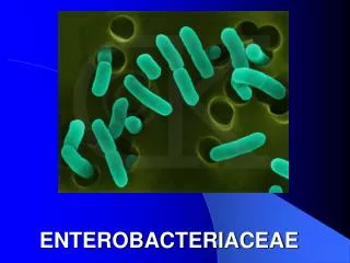
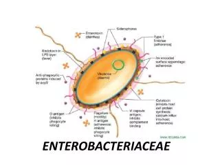
Enterobacteriaceae. Escherichia. E. coli Theodore Escherich in 1885 most significant species common isolate from the colon flora black/green metallic sheen on EMB posesses O ( Shigella ), H and K (Group B N. meningitidis ) Ag.

E N D
Escherichia • E. coli • Theodore Escherich in 1885 • most significant species • common isolate from the colon flora • black/green metallic sheen on EMB • posesses O (Shigella), H and K (Group B N. meningitidis) Ag. • other species: blattae, vulneris, fergusonii, hermanii
Diarrheal Infections* EPEC – enteropathogenicE. coli - infantile diarrhea - watery stool w/ mucus but w/o blood* ETEC – enterotoxigenicE. coli - traveller’s diarrhea - infective dose of 106- produce LT and ST toxin (hypersecretion) - non-bloody, watery diarrhea with abdominal cramps and low grade fever
Diarrheal Infections • EIEC – entero invasive E. coli • cause dysentery ( direct penetration, invasion and destruction of the intestinal mucosa) – similar with dysentery • scanty stool with pus, mucus and blood • Sereny test – determines the invasiveness of EIEC • (+) result – keratoconjunctivitis in guinea pig
Diarrheal Infections EAEC – enteroadherent E. coli cause diarrhea by adhering to the mucosal surface of intestine. watery diarrhea, vomiting and dehydration EHEC – enterohemorrhagic E. coli or VTEC (E. coli 0157:H7) Hemorrhagic diarrhea, colitis and hemolytic uremic syndrome (HUS) Bloody diarrhea and crampy abdominal pain
Microscopic Exam E. coli
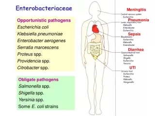
Other Infections • Septicemia and Meningitis • accounts for 40% of neonatal meningitis • Urinary Tract Infections • 90% of cases of UTI
Shigella 4 subgroups Fermentation of lactose mannitol ODC ONPG A S. dysenteriaenegnegnegneg B S. flexnerinegposnegneg C S. boydiinegposnegneg D S. sonneinegpospospos
Shigellosis or Bacillary Dysentery watery diarrhea bloody stool with WBC’s & mucus fecal-oral route (human – only known reservoir) self-limiting but highly communicable bec. of low infective dose (200 bacilli) Gay bowel syndrome (S. flexneri) Clinical Infections - Shigella
Edwardsiella • Edwardsiella • tarda (human pathogen – bacteremia and wound infection) • hoshinae (snakes, birds and water) • ictaluri (enteric septicemia in fish)
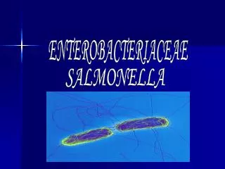
Salmonella • Salmonella • 2200 species • 3 common species (cholerasuis, typhi, enteritidis) • 7 subgroups (with Arizona as groups 3A & 3B) • Virulence factors • O, H and Vi antigens
Salmonella • Gram negative rods • Do not ferment lactose • Produce H2S • Major cause of food poisoning in the U.S. • Cause intestinal infections (enterocolitis); enteric fevers (typhoid fever); and systemic infections (septicemia) • Chickens are a major reservoir for this bacteria • Identification on basis of antigens (O, H, Vi)
Salmonella - pathogenesis Enterocolitis: invasion of epithelial cells of intestines infectious dose (ID50) ~ 100,000 organisms infection of gut - inflammation and diarrhea, self correcting Typhoid: starts in small intestine: enters and multiplies in mononuclear cells; spreads to liver, gallbladder, spleen. Leads to bacteremia with onset of fever. carrier state - organisms excreted in feces Septicemia: minority of infections, usually in patients with underlying disorder
S. enteritidis S. typhimurium S. typhi
Citrobacter • resembles Salmonella but are ONPG (+) & LDC (-) • Citrobacter species • freundii(UTI, pneumonia, intraabdominal abscess) • diversus (neonatal meningitis) • amalonaticus (extraintestinal infections) C. freundii S. diversus H2S + - Indole - + KCN + -
Klebsiella Klebsiella-Enterobacter-Serratia-Hafnia K. pneumoniae >Friedlander’s bacilli (encapsulated and mucoid) > (+) String test K. oxytoca – similar w/ K. pneumoniae except for its indole production K. ozanae– from nasal secretions K. rhinoschleromatis– rhinoscleromatis (infection of nasal cavity with intense swelling and malformation of the entire face and neck)
Enterobacter • species: habitat: soil, water, dairy products • normal flora of the git of animals & humans • motile, ODC & ONPG (+)
Enterobacter LDC ADH ODC Urease Yellow pigment • E. cloacae - + + V - • E. aerogenes + - + - - • E. gergoviae + - + + - • E. sakazakii - + + - + • E. taylorae - + + - -
Serratia • opportunistic pathogens • DNAse, lipase, gelatinase • S. marcescens • S. liquefaciens • S. rubidaea • S. oderifera
Hafnia • Hafniaalvei • 2 biotypes • H. alvei • H. alvei Biotype 1 – associated in breweries • delayed (+) citrate reaction is its major characteristic
Proteus • rapid urease producers • Swarming, burned chocolate odor P. mirabilis P. vulgaris Indole - + ODC + - Fermentation • Maltose - + • Xylose + + • Salicin - + Chloramphenicol S R
Proteus mirabilis Swarming pehomenon Proteus
Morganella • Formerl known as Proteus • M. morganii – only specie • UTI’s and wound infections
Providencia Providencia ( 4 species – P. alcalifaciens, P. rustigianii) P. rettgeri P. stuartii Urease + - Citrate + +
Yersinia • 11 species • Y. pestis (plague) – bipolar staining (wayson) • Bubonic – bite of infected flea • Pneumonic • Prefered growth at 25 deg C • Y. enterocolitica (acute enteritis – appendictis like) • Pig, cats and dogs • Cold enrichment • Motile at RT • Y. pseudotuberculosis • Pathogen in animals (turkey, geese, doves, farm and domestic animals)
Agent of bubonic plague, pneumonic and septicemic plagues • Bubonic plague contracted via flea bites • Y. pestis sheds capsule while growing in invertebrate host. • In humans most of the microbe is taken up and killed by PMN cells • Some organisms taken up by Macrophages which can’t kill pestis. • Organism multiples in Mac and resynthesizes capsule • Organism kills Mac and is released into extracellular environment. • The encapsulated microbe resists phagocytosis and spreads to lymph nodes which become swollen, and hemorrhagic giving the black buboes characteristic of the disease. • Microbe rapidly spreads through blood, liver, spleen, lung giving rise to highly contagious pneumonia (highly fatal).
Yersinia pestis Yersinia Safety pin appearance
Erwinia • Plant pathogens • Fail to grow in CM used in the isolation of enteric bacilli