Download
1 / 61
1.1k likes | 2.57k Views
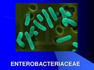
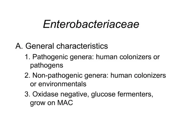
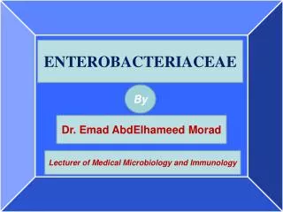
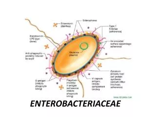
Enterobacteriaceae. Dr . Neha Haswani Assistant Professor, Dept .of Microbiology. Common properties. Inhabitants of intestine of humans &animals Grows on macconkeys agar Aerobic & facultative anaerobes Catalase positive Oxidase negative Reduce nitrates to nitrites.

E N D
Enterobacteriaceae Dr.NehaHaswani Assistant Professor, Dept .of Microbiology
Common properties • Inhabitants of intestine of humans &animals • Grows on macconkeys agar • Aerobic & facultative anaerobes • Catalase positive • Oxidase negative • Reduce nitrates to nitrites
Differentiating features • Lactose fermentation • H2 S production • Motility • Sugar fermentation tests • Gas production • IMViC reactions- Indole, Methyl red, Vogesproskauer, Citrate • Urease production • Decarboxylation of lysine & ornithine • Deamination of lysine • Dehydrolisation of arginine • Pigment production
Growth on macconkey agar Differential medium
Indole test Methyl red(MR) IMViC tests Voges proskauer(VP) Citrate test
TRIPLE SUGAR IRON AGAR A B C D
Classification • Tribe-I Escherichiae • Escherichia • Shigella • Tribe-II Klebsiellae • Klebsiella • Enterobacter • Hafnia • Serratia • Tribe-III Proteeae • Proteus • Morganella • Providencia • Tribe-IV Erwinieae • Erwinia • Tribe-V Erdwardsiella • Erwinia • Tribe VI Citrobacteriaceae • Citrobacter • Tribe-VII Salmonellae • Salmonella • Tribe-VIII Yersinieae • Yersinia
Escherichia • Escherichia coli • Escherichia fergusonii • Escherichia hermanii • Escherichia vulneris • Escherichia blattae
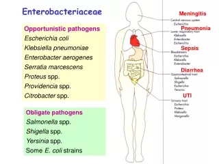
Escherichia coli Urinary tract infection Diarrhoea Pyogenic infections Septicaemia & meningitis (neonates)
Pathophysiology • Bacterial virulence factors: • Lipopolysaccharide • Fimbriae : ‘P’ fimbriae (to uroepithelial cells) • Enterotoxins – 3 types • Bacterial invasion • Colonisation factor antigens • Host factors • Urinary obstruction • catheterisation • Age • Travel UTI Diarrhoea Anatomical anomalies Prostatic hypertrophy Pregnancy calculi
Urinary tract infection • Definition :UTI is defined as bacteriurea i.e. the multiplication of bacteria in urine with in the renal tract. Significant bacteriuria : KASS concept In symptomatic patients if >105 CFU of single strain/1 ml of mid stream urine sample is isolated, it represents significant bacteriuria
Significant bacteriuria Population Significant bacteriurea Asymptomatic bacteriuria Acute pyelonephritis Women with acute dysuria Patients with indwelling catheter >105 CFU /ml > 105 CFU/ml > 102 CFU/ml > 102 CFU/ml
Classification Based on anatomical site Based on epidemiology Lower UTI Urethritis Cystitis Prostatitis Upper UTI Pyelonephritis Intrarenal &perinephric abscesses Nosocomial UTI Community acquired UTI
Causative agents Un complicated Complicated Enterobacteriaceae members: E . coli , Klebsiella ,Proteus & others S .aureus S .saprophyticus Enterococci Group B Streptococci Chlamydiae Pseudomonas aeruginosa S .epidermidis Enterobacteriaceae
Risk factors for UTI • Structural abnormalities that obstruct the flow • Indwelling catheter • Diabetis mellitus • Immunosuppressed patients • Reflux of urine in children • Sexual intercourse in females • Contraceptive devises • Neurogenic bladder/bladder diverticulum • Post menopausal women with bladder or uterine prolapse • Pregnant women
Clinical features • Fever with chills and rigor • Burning micturition/ dysurea • Urgency & increased frequency with small volume of urine voiding • Tenderness in flank • Supra pubic tenderness • Nausea & vomiting • Turbid or cloudy urine
Lab diagnosis • Specimen collection : • Clean catch mid stream urine (10-20ml) • Catheterised urine sample • Supra pubic cystostomy sample • Specimen should be processed with out delay • Preservation : (up to 4 hrs ) Refrigeration Preservatives like boric acid , sodium formate
Microscopy & Culture • Wet mount • Gram’s stain • Culture – semiquantitative culture Calibrated loop method Filter paper strip method • Rapid diagnostic techniques
4mm diameter loop (0.01ml of urine) Semiquantitative culture by calibrated loop method No of colonies x 100 = colony count
Macconkey ‘s agar Lactosefermenting colonies on macconkey agar
CLED MEDIUM(CYSTEIN LACTOSE ELECTROLYTE DEFICIENT MEDIUM)
Identification • Lactose fermenting ,low convex,smooth colonieswith regular margin • Motile • Ferments sugars ( Glu, lac,mannitol) with acid &gas • Urease negative • IMViC= + + - -
Screening tests for UTI • Griess nitrite test • Catalase test • Triphenyltetrazolium chloride test • Gram’s stain • Glucose test paper • Dipslide culture methods
DIARRHOEA • Entero toxigenic E. coli (ETEC) • Enteropathogenic E. coli (EPEC) • Enteroinvasive E. coli (EIEC) • Enterohemorrhagic E. coli (EHEC) • EnteroaggregativeE. coli (EHEC) • Diffusely adherent E. coli (DAEC)
Enterotoxigenic E.coli (ETEC) • Endemic diarrhea in infants in developing countries • Traveller’s diarrhea • Enterotoxin mediated • LT (cholera like toxin)- heat labile enterotoxin • ST- heat stable enterotoxin • S erotypes: O6,8,15,25,27,63,78,115,148,153,159 • Resembles cholera • Noninvasive, toxigenic • Pathogenesis – production of plasmid coded toxins(LT/ ST) • Clinical features - Diarrhea, vomiting and abdominal pain • Lab Diagnosis – demonstration of enterotoxin by in vitro or in vivo methods, detection of LT/ St by gene probes
Enterotoxigenic E. coli (ETEC) LT ST • Toxigenicity tests : • In vivo : Rabbit ileal loop + + Vascular permeability + _ • In vitro: Tissue culture + _ • ELISA
Enteropathogenic E.coli (EPEC) • Infantile diarrhea • Institutional outbreaks • Noninvasive, nontoxigenic • Pathogenesis – adhesion via fimbria, disruption of brush border microvilli • Serotypes : O111, O26, O55 • Clinical features – fever, diarrhea, vomiting, nausea, non bloody stools • Lab Diagnosis – testing colonies grown on BA/ MA with EPEC O antisera
Enteroinvasive E.coli (EIEC) • Bloody diarrhea (dysentery), resembles Shigella dysentery • Passage of blood, mucus & leucocytes in stool • Pathogenesis - Invades epithelial cells by endocytosis and can spread laterally to adjacent cells, causes tissue destruction, necrosis and ulceration. • Lab Diagnosis: • Nonmotile • Non lactose fermenting colonies • No gas • LIA – decarboxylation positive • Serotyping • Sereny test- instillation of suspension of freshly isolated EIEC or Shigella in the eyes of guinea pig – mucopurulent conjunctivitis and severe keratitis • Penetration of HeLa or Hep2 cells in tissue culture
Enterohemorrhagic E.coli (EHEC) • Produces verocytotoxin (VT), a shiga-like toxin (SLT); hence also known as Verocytotoxigenic E.coli (VTEC) • Pathogenesis – EHEC attaches to the colonic mucosa and releases VT. It targets vascular endothelial cells, inhibits protein synthesis - cytotoxicity • Clinical features - Mild diarrhea (bloody) to fatal complications (esp. in young children and elderly): • Hemorrhagic colitis – destruction of mucosa followed by hemorrhage. • Hemolytic Uremic syndrome – triad of acute renal failure, hemolytic anemia and thrombocytopenia. • Serotype O157: H7 is most commonly involved. • Outbreaks of food poisonings (fast foods, contaminated hamburgers)
Enterohemorrhagic E.coli (EHEC) • Lab Diagnosis: • Demonstration of bacilli or VT in feces or in culture • Sorbitol MacConkey agar for O157:H7 – does not ferment sorbitol unlike other E.coli • Cytotoxic effects on Vero or HeLa cells • DNA probes to detect toxins
SORBITOL MACCONKEY AGAR O157:H7
Enteroaggregative E.coli (EAEC) • Persistent diarrhea in children in developing countries. • Aggregate to give a “Stacked brick appearance” on Hep2 cells or glass (due to fimbria) • Pathogenesis – shortening of villi, mucus biofilm, heat stable cytotoxin (hemorrhagic necrosis and edema)
Epidemiology & Treatment • EPEC & ETEC - most important causes of diarrhea globally • EHEC – in developed countries. Epidemiology Treatment • Based on symptoms: • Primary treatment – fluid replacement • Secondary treatment – antibiotics in severe cases with systemic involvement
Klebsiella Non motile Capsulated Lactose fermenting bacilli
K.Pneumoniae • Pneumonia • UTI (usually catheter related) • Pyogenic infections • Septicaemia • K.Ozaenae • Ozena • K. Rhinoscleromatis • Rhinoscleroma • K. Oxytoca • Neonatal septicaemia/bacteraemia • Wound infection
Klebsiellapneumoniaeis the most important species • 2ndmost abundant organism seen in intestine of humans • One of the common organism associated with HAI • Gram negative • Non motile • Nonsporing • Capsulated- polysaccharide, major virulence factor • Produces large dome shaped mucoid colonies
Pneumonia • Middle age or old people • Risk factors for pneumonia • Alcoholism • Chronic bronchopulmonary disease • Diabetes mellitus • Characteristic features seen in clinical disease • Destructive in nature • Mucoid sputum • Involves one or more lobes • Necrosis and abscess formation is frequent • Serotypes 1,2,3 are commonly involved
Also causes pyogenic infection like abscesses, meningitis, septicaemia • Neonatal cases associated with high mortality • Treatment is a serious problem • As majority strains are MDR (plasmid mediated) • .
Diagnosis • Culture and sensitivity • Lactose fermenting ,mucoid colonies • Capsulated short plump bacilli • Non motile • Ferments sugars ( Glu, lac, suc,mannitol) with acid &gas • Urease positive • IMViC= - - + +
Enterobacter • Lactose fermenter • Resembles Klebsiella • Motile • Normally found in sewage,soil, feces,water • E. cloacae, E. aerogenes – two clinically relevant species • Causes UTI & septicemia • Usually responsible for HAI
HAFNIA • Only 1 species- H.alvei. • Found in human and animal faeces, soil, sewage and water. • Rare in clinical specimens • Motile • Non lactose fermenting colonies
SERRATIA • Characterised by production of Red or Pink coloured non diffusible pigment- Prodigiosin • Only one species is medically important- Serratiamarcescens • It is pleomorphic coccobacilli • Saprophyte found in water, soil and food. • Increasingly associated with healthcare associated infection • Contaminates iv fluids, surgical instruments antiseptic solution • Causes meningitis, septicaemia, respiratory infections. • MDR commonly seen.
Tribe Proteae Proteus Providencia Morganella
Common features • Part of commensal in intestine • Fishy odour • Motile • Non lactose fermenter • Urease positive • PPAtest – positive
PROTEUS • Gram negative • Pleomorhic • Motile • Non capsulated • Saprophytes-found in decoposing animal matter,sewage soil • Comensal in the gut. • Two important spp-Pr.mirabilis & Pr. Vulgaris • Other less commonly isolated spp are Pr.penneri & Pr.myxofaciens
Opportunistic pathogens • UTI • Wound &soft tissue infection • septicaemia • Associated with struvite stones • Forms the basis of Weil Felix test- A Heterophile agglutination test for diagnosis pf rickettsial infections.