Download

1 / 63
640 likes | 840 Views
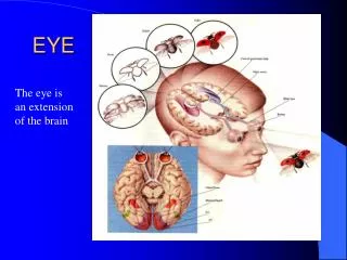
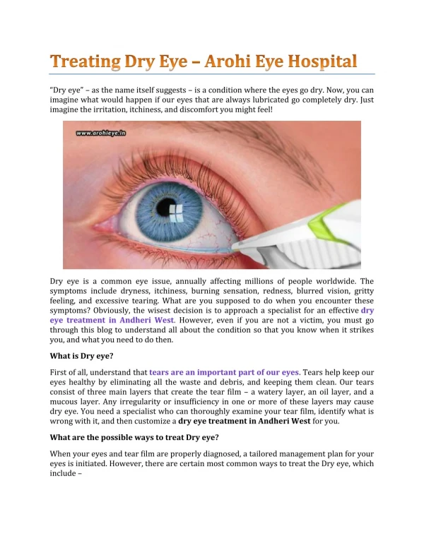
EYE. Jittipan Chavadej,Ph.D. Microanatomy for Medical students. Dept. of Anatomy, Fac. of Science Mahidol University. Nov. ,43. Transverse section of the eye. Structure of the eye. Hollow, spherical structure dia.~2.5 cm 3 distinct layers

E N D
EYE Jittipan Chavadej,Ph.D. Microanatomy for Medical students Dept. of Anatomy, Fac. of Science Mahidol University Nov. ,43
Structure of the eye • Hollow, spherical structure • dia.~2.5 cm • 3 distinct layers -outer fibrous tunic (corneoscleral coat) -middle vascular tunic(choroid) -inner nervous tunic(retina)
Sclera • -thick, fibrous layer~ 0.6-1mm thickness • collagen,elastic & fibroblasts(betw. fiber bundles) • Tenon’s capsule-thin sheet of CNT separating eyeball from adipoes tissue • Episcleral space-betw sclera&Tenon’s capsule--> rotation of eyeball
Cornea Corneal epithelium ~5 layers of unkeratinized cells, rich insensory nerve ending, regeneration Bowman’s membrane -lamina 10-12um thick -thin collagen fibrils(random) Stroma (substantia propria) -90% of the thickness of cornea -lamellae of collagen fiber(200-250) -fibroblast&fiber-chondroitin sulphate and keratan sulphate
Endothelium -simple squamous epi. -inner surface of cornea -numerous pinocytotic vesicles -Na+ pump-> Na+ into antr chamber =resorption of excess fluid->dehydration of stroma Descemet’s membrane -thick basement membrane-underlying endothelium -thin atbirth-->older-thicker (thin collagen fibrils)
Sclerocorneal junction(limbus) -transition betw sclera & cornea -inner surface-trabecular meshwork strands of CNT (covered by endothelium) -->Schlemn’s canal (tubular space -> deep vein of the limbus -> epicleral vein) -drainage pathway of aqueous humor
Clinical correlation • Glaucoma -failure of drainage of aq. humor --> increased intraocular pressure -lead to progressive damage to the eye, retina --> blindness (untreated) Drawing of sclerocorneal junction,iris, lens, and ciliary body as seen in meridian section
Middle layer: Tunica vasculosa -is composed of 3 parts 1. Choroid 2. Ciliary body 3. Iris
Choroid -thin, highly vascular layer -contain many blood vv - loose CNT -black color-presence of melanocytes -inner surface-choriocapillary layer for providing nutrient to retina -Bruch’s mem.-refractile layer -antrend-ora serrata(dentated antr margin of retina)
Ciliary body • thickening of vascular tunic • epith.-2layers+ loose CNT Epithelium- 2layers -outer=unpigmented -inner=pigmented Unpigmented epith.:mitochondria, RER etc Zonule fibers-fibrillin :--> lens for anchoring lens in place • Ciliary processes: 70 processes radiate out from the central core (fenestrated-capillaries) • folding of plasma mem.-transporting H2O,ions-aq. humor
SEM micrograph of postr surface of the lens. note:lens, suspensory ligament, ciliary body
Ciliary muscle 2 bundles of sm. muscle -is inserted into sclera -is inserted along the inner wall of ciliary body *Contraction-release tension --> lens : more convex Contraction of ciliary muscle Relaxation of ciliary muscle
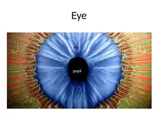
Iris:antr extension of choroid --> pupi -stroma: CNT+melanocyte -antr surface:incomplete layer -postr surface:2layers of epith. *Dilator pupillae muscle *Sphinctorpupillae muscle
Refractive media of the eye : • cornea • lens • vitreous body Vitreous body -transparent refractile gel -adhere to the retina over the entire surface -99%=H2O+electrolyte+collagen fibers+hyaluronic acid
Lens -flexible, biconvex -consist of 3 parts -lens capsule -subcapsular epithelium (antr surface of the lens) -lens fibers-2000 fibers 7-10mm long -mature crystallins, few organelles EM micrograph of lens fibers
Macula lutea -2.5mm lateral to the optic disk -1.5mm in diameter -fovea centralis: 500um *greatest visual acuity *only cones are present Blind spot:optic disk =exit site of the optic nerve
Optic disk(optic papilla) -small round area -devoid of photoreceptor cells Retina seen through the pupil showing optic disk and macula lutea(yellow spot) & fovea centralis
Retina 1 note: sclera,choroidretina 4 6 8 10 distinct layers 1.pigment epithium 2.layer of rods & cones 3.outer limiting mem. 4.outer nuclear layer 5.outer plexiform layer 6.inner nuclear layer 7.inner plexiform layer 8.ganglion cell layer 9.optic nerve fiber layer 10.inner limiting mem.
Pigmented epithelium Outer nuclear layer -nuclei of rods & cones Inner nuclear layer -nuclei of bipolar neurons -amaccrine,horizontal, Muller cells Ganglion cell layer -multipolar neurons
2-3Rods 2-3bipolar neurons 1ganglion cell-->Single nerve fiber 1 Cone 1 bipolar neuron 1 ganglion cell
Rod:activated in dim light • Outer segment • several hundred flattened membranous lamellae(invagination of plasma mem.-->detached from cell surface) • membrane contain rhodopsin (a light-sensitive pigment) • Inner segment • mitochondria-glycogen granules RER,SER,golgi cpx. • proteins-->outer seg.->disk
Cone :activated in brightlight • Lamellae-attached to plasma membrane • are sensitive to color
Diagram of the morphology of a rod and cone note:outer&inner seg., connecting stalk, synaptic region
EM micrograph of portion of outer & inner segment of cone Note:ellipsoid-shape mitochondria EM micrograph of portion of outer & inner segment of rod
Pigment Epithelium -cuboid to columnar cells -mitochondria-cell invagination -*desmosome-zonulae occludentes-zonulae adherentes--> lateral cell mem.-forming blood-retina barrier -apical surface->microvilli & sleeve-like structure-surround the tip of rod & cone cells (detached retina-hard jolt)
Pigment epithelium(cont.) -abundance of melanin granules -function:absorb light after it has passed through and stimulated the photoreceptor cells-->preventing reflections from the tunic phagocytized spent membranous disks from the tip of the rods play an active role in vision by esterifying vit.A derivatives in their SER.
Eyelids -skin -loose CNT-muscles -tarsal plate-dense CNT Meibomian gland(modified sebaceous gland)->thin lipid layer over tear film-retard its evaporation
Lacrimal glands -tubuloalveolar gland -alveoli-serous columnar cells -tear film -over the bulbar conjunctiva and cornea -tear--> lacrimal puncta-->lacrimal duct-->lacrimal sac-->nasolacrimal duct-->infr meatus of nasal cavity
EAR Jittipan Chavadej, Ph.D. Dept. of Anatomy,Fac. of Science, Mahidol University Microanatomy for Medical student Dec.43
3 regions : • external ear, • middle ear, and • internal ear • External ear • Auricle: elatic cartilageplate-skin • External auditory meatus:skin lining +cerumenous gland (modified sweat gland) -->tympanic membrane
Middle ear Tympanic cav.: Air-filled space Ear ossicles Tensor tympani Stapedius Eustachain tube • Tympanic membrane: -outer surface-thin layer of epidermis-middle-tough layer of CNT-inner surface-sq. epith. that line tymp. cavity
Tympanic cavity communicates : -antr-->nasopharynx-Eustachain tube -postr --> mastoid air cells -oval&round windows-medl wall, -connect to inner ear Ear ossicle chain : -tympanic membrane - malleus--incus-->stapes - oval window
Middle ear -2 small muscles • tensor tympani-medl surface of malleus & cartilagenous wall of auditory tube-->pull malleus inward • stapedius-postr surface of stapes&inner wall of tympanic cav.-->pull stapes outward • Loud sound-->contraction of mus.->movement of malleus & stapes • bridge of ear ossicles-more rigid->vibration to inner ear is reduced
Auditory tube • Connect the middle ear to the throat • Helps to maintain equal air pressure on both sides of the ear drum • Usually closed-open by swallowing, yawning-->hasten the equalization of air pressure during altitude changes • Provide the route for middle ear infection
Inner ear • Osseous labyrinth -bony canal • memb. labyrinth -tube lies within the bony canal A B A=bony labyrinth B=bony cochlea-mem. part C=membranous labyrinth C
Inner Ear • Vestibule- Utricle and saccule • Semicircular canals -Semicircular ducts • Cochlea- Cochlear duct
Perilymphatic space-betw. bony & memb. parts - perilymph • perilymph-is secreted by the cells in the wall of the bony canal • All portions of membranous labyrinth contain a fluid = endolymph
Vestibule - saccule & utricle • Wall-thin outer vascular leyer of CNT • -inner layer of simple squamous-low cuboidal epithelium • specialized region=receptors saccule-macula sacculi -floor utricle-macula utriculi -lateral wall
M. utriculi/ sacculi • Hair cells • otolithic membrane • otolith
Type I Hair Cell • Round base,narrow neck • RER,Golgi cpx,numerous small vesicles • Single kinocilium • Stereocilia-dense terminal web(actin filaments cross-linked by fimbrin) bending at the neck region • surrounded by a cup-shaped afferent nerve fiber
Type II Hair Cell • Similar to type I hair cell • longer,more columnar in shape • many small nerve endings • synaptic ribbons-function in synapses with efferent nerves for modulaing sensitivity
B A SEM micrograph of the bundle of stereocilia on the vestibular hair cell A-hair cell of bull frog b-hair cell found in utricle & saccule
Supporting cell • A few microvilli-apical surface • a well-developed Golgi cpx.+ secretory granules • thick junctional cpx. to the hair cells • fn.:-help to maintain hair cell ? -may contribute to the production of endolymph ?(dark cell of non receptor region)
Otolithic membrane : • thick gelatinous glycoprotein mass • otolith (otoconia)-on the surface region of otolithic membrane • otolith=small calcium carbonate crystals
Bending the head - cause displacement of endolymph-->otolith->bending of stereocilia->action pot.->nerve impulse->brain-inform the position of the head
Semicircular ducts • 3 ducts-perpendicular to each other • lined by cuboid epithelium • contain endolymph • each has a small dilatation=ampulla • transverse ridge(floor of ampulla)=crista ampullaris-receptor region