Download
1 / 19
210 likes | 286 Views
Learn about outer retinal tubulation (ORT) identified in a case of choroidal neovascular membrane, its significance in diagnosis, treatment, and associated retinal disorders. Explore images and findings from optical coherence tomography scans.

E N D
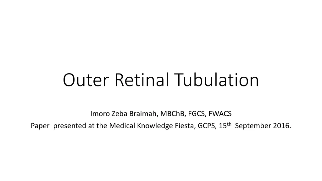
Outer Retinal Tubulation Imoro Zeba Braimah, MBChB, FGCS, FWACS Paper presented at the Medical Knowledge Fiesta, GCPS, 15th September 2016.
INTRODUCTION • SPECTRAL DOMAIN OPTICAL COHERENCE TOMOGRAPHY
INTRODUCTION • Branching tubular structures- outer nuclear layer of retina • round or ovoid hyporeflective spaces with hyperreflective borders on SD-OCT. SD-OCT= spectral-domain optical coherence tomography, ORT=outer retinal tubulation, CME= cystoid macular edema Image courtesy of Sandrine A. Zweifel 1. Zweifel SA, Engelbert M, Laud K, et al: Outer retinal tubulation: a novel optical coherence tomography finding. Arch Ophthalmol 2009; 127: 1596–1602.
Case • 66 years, Male with Choroidal Neovascular membrane both eyes-5yrs duration. • Came for follow up – April 2016 • Had monthly injections of intravitreal anti-vascular endothelial growth factors (anti-VEGF) agents in right eye • bevacizumab-12 injections • Ranibizumab-6 injections.
Past History • First seen in KBTH-16/05/2014- sudden painless loss of vision Right eye, micropsia, metamorphopsia and inability to read– 2 days duration. • Had diabetes mellitus and hypertension under control
C A B Fundus Photograph and SD-OCT at presentation E D F
Diagnosis and Treatment • Diagnosis- Choroidal neovascular membrane(CNVM) Both eyes. Right active, left eye fibrotic. • Plan of treatment • Intravitreal injection of anti-VEGF right eye • Observe left eye
B A C F E D Images A,B,C- Shows SD OCT Scans of the right eye at presentation and Images D, E, and F- shows the SD OCT scans after 3 injections of intravitreal bevacizumab in same eye
15 Months after Initial Bevacizumab Injection • Acuity right eye 6/18 • Intravitreal bevacizumab stopped and Ranibizumab started September 2015
24 Months after Initial Bevacizumab Injection • SD-OCT of both eyes with ORT in the left eye
27 Months after Initial Bevacizumab Injection SD-OCT Right eye showing new onset PED and Left Eye showing ORT
29/08/2016 28 months SD-OCT RIGHT EYE WITH HEMORRHAGIC PED AND LEFT EYE WITH ORT
Diagnosis • Right Eye Choroidal neovascular membrane(CNVM)with persistent Intraretinal fluid and pigment epithelial detachment • Left Eye fibrotic CNVM with OUTER RETINAL TUBULATION
OUTER RETINAL TUBULATION(ORT) • Occur due to rearrangement of degenerating/surviving photoreceptors • Advanced degenerative retinal disorders. Image courtesy of Sandrine A. Zweifel 1. Zweifel SA, Engelbert M, Laud K, et al: Outer retinal tubulation: a novel optical coherence tomography finding. Arch Ophthalmol 2009; 127: 1596–1602.
DISEASE ASSOCIATIONS • Neovascular AMD(nvAMD) • Gyrate atrophy • Pseudoxantoma elasticum • Bietti crystalline dystrophy • Pattern dystrophy • Stargardts disease • Retinitis pigmentosa • Best Disease • Adult vitelliform foveomacular dystrophy Zweifel et al 2009 Goldberg et al 2013 Iriyama et al 2013 Dolz-Marco et al 2013 Braimah et al 2016
Clinical Significance of ORT • May simulate Cystoid macular edema and subtretinal fluid • Associated with poor visual acuity outcome in nvAMD • Indicator of disease severity Lee et al 2014 Faria- Correia et al 2013 Espina et al 2015 Goldberg et al 2013
Composite image of the right eye of a 32-year-old man with Best disease with stable ORT over a follow-up of 3 years
CONCLUSION • ORT was identified in the left eye with fibrotic CNVM and worse visual acuity. • It is important to identify ORT in eyes with CNVM to avoid unnecessary treatment