Download
1 / 51
580 likes | 806 Views
This guide covers the definitions, anatomy, examination techniques, and pathogenesis of retinal detachment (RD), including retinal breaks, morphology of tears, normal anatomical landmarks, examination methods like indirect ophthalmology and scleral indentation, and types of RD like rhegmatogenous, non-rhegmatogenous, and diabetic tractional RD. Learn about the classification of RD, signs to look for in fresh and longstanding RD, and the pathogenesis of conditions like proliferative vitreoretinopathy (PVR) and exudative RD.

E N D
Dr Seemant Raizada MBBS, MS, DNB, FRCSEd Vitreo-Retinal Surgeon Retinal Detachment
INTRODUCTION TO RETINAL DETACHMENT (RD) 1. Definitions and classifications • Retinal breaks • Retinal detachment 2. Anatomy • Anatomical landmarks • Variants of ora serrata • Vitreous base 3. Examination techniques • Indirect ophthalmology • Scleral indentation • Fundus drawing • Slitlamp biomicroscopy
Definition and classification • Break - full-thickness defect in sensory retina • Hole - caused by chronic retinal atrophy • Tear - caused by dynamic vitreoretinal traction Morphology of tears a. Complete U-tear b. Linear c. Incomplete L-shaped d. Operculated e. Dialysis
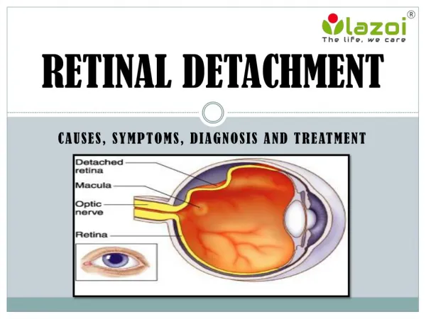
Retinal detachment (RD) Separation of sensory retina from RPE by subretinal fluid (SRF) Rhegmatogenous - caused by a retinal break Non-rhegmatogenous - tractional or exudative
Normal anatomical landmarks Short ciliary arteries Temporal ora serrata Nasal ora serrata Short ciliary nerves Vortex ampullae Macula Long ciliary artery Long ciliary nerve Microcystoid degeneration Vortex vein Short ciliary nerves
Normal variants of ora serrata a. Meridional fold a b c • Small radial fold in line with • a dentate process • Occasionally small hole at base b. Enclosed oral bay • May be mistaken for retinal • hole c. Granular tissue • Multiple, tiny, white opacities • May be mistaken for small • opercula
Anatomy of vitreous base Pars Plicata Pars Plana Vitreous base • 3-4 mm wide zone straddling ora serrata • Strong adhesion of cortical vitreous • Anterior limit of posterior vitreous detachment
Indirect ophthalmology Condensing lenses Technique • Keep lens parallel to patient’s iris plane • Avoid tendency to move towards patient • Ask the patient to move eyes and head • into optimal positions for examination • The higher the power, the less the • magnification, the shorter the working • distance but the greater the field of view
Scleral indentation Retinal breaks in detached retina without indentation Enhanced visualization of breaks with indentation
Fundus drawing Technique Colour code Breaks Detached retina Vitreous opacity Thinning Exudate Lattice Retinal pigment • Place chart upside down • Draw what you see
Slitlamp biomicroscopy Goldmann triple-mirror lens View of peripheral fundus • Image is upside down • Equatorial mirror (largest and • oblong) - from 30 to equator • Peripheral mirror (square) - • from equator to ora serrata • Gonioscopic (smallest)
Primary retinal break It is responsible for RD and determines configuration of SRF Quadratic distribution of breaks in eyes with RD Configuration of SRF in relation to primary break
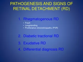
PATHOGENESIS AND SIGNS OF RETINAL DETACHMENT (RD) 1. Rhegmatogenous RD • Fresh • Longstanding • Proliferative vitreoretinopathy (PVR) 2. Diabetic tractional RD 3. Exudative RD 4. Differential diagnosis RD
Pathogenesis of rhegmatogenous RD • Two components for retinal break formation • Acute posterior vitreous detachment (PVD) • Predisposing peripheral retinal degeneration Possible sequelae of acute PVD Retinal tear formation and haemorrhage (10-15%) Avulsion of retinal vessel and haemorrhage (uncommon) Uncomplicated PVD (85%)
Fresh rhegmatogenous RD - signs • Annual incidence - 1:10,000 of population • Eventually bilateral in 10% • Loss of choroidal pattern • Retinal breaks • Convex, deep mobile elevation • extending to ora serrata • Slightly opaque with dark blood vessels
Longstanding rhegmatogenous RD - signs • Frequently inferior with small holes • Very thin retina • Secondary intraretinal cysts • Demarcation lines (high-water marks)
Classification Of PVR • The term "proliferative vitreoretinopathy" was coined in 1983 by the Retina Society Terminology Committee. • In 1989, the classification was amended by the Silicone Study Group before being most recently modified in 1991 to its current classification. • Currently, PVR is divided into grades A, B, and C. • Grade A is limited to the presence of vitreous cells or haze. • Grade B is defined by the presence of rolled or irregular edges of a tear or inner retinal surface wrinkling, denoting subclinical contraction. • Grade C is recognized by the presence of preretinal or subretinal membranes. Grade C is further delineated as being anterior to the equator (grade Ca) or posterior to the equator (grade Cp) and by the number of clock hours involved (1 to 12).
Proliferative vitreoretinopathy Grade A (minimal) Grade B (moderate) Grade C (severe) • Vitreous haze and • tobacco dust • Retinal wrinkling and • stiffness • Rolled edges of tears • Rigid retinal folds • Vitreous condensations • and strands
Pathogenesis of diabetic tractional RD (1) Antero-posterior traction RD Preretinal haemorrhage
Pathogenesis of diabetic tractional RD (2) A-P traction Bridging traction Preretinal haemorrhage
Signs of diabetic tractional RD • Slow progression and variable fibrosis • Does not extend to ora serrata • Concave, shallow immobile elevation • Highest at sites of vitreoretinal traction
Pathogenesis and Causes of Exudative RD • Damage to RPE by subretinal disease • Passage of fluid derived from choroid into subretinal space 1. Choroidal tumours • Primary • Metastatic 2. Intraocular inflammation • Harada disease • Posterior scleritis 3. Systemic • Toxaemia of pregnancy • Hypoproteinaemia 4. Iatrogenic • RD surgery • Excessive retinal photocoagulation 5. Miscellaneous • Choroidal neovascularization • Uveal effusion syndrome
Signs of exudative RD • Convex, smooth elevation • May be very mobile and deep with • shifting fluid • Subretinal pigment (leopard spots) • after flattening
Differential diagnosis of RD Degenerative retinoschisis Choroidal detachment Uveal effusion syndrome • Frequently bilateral • Smooth, thin and immobile • Occasionally breaks in one • or both layers • Associated with hypotony • Unilateral, brown, smooth, • solid and immobile • Ora serrata may be visible • Idiopathic • Rare, unilateral • Combined choroidal and • exudative detachments
PROPHYLAXIS OF RHEGMATOGENOUS RETINAL DETACHMENT 1. Retinal breaks 2. Predisposing degenerations • Lattice • Snailtrack • White-without-pressure 3. Treatment modalities • Laser photocoagulation • Cryotherapy 4. Benign peripheral degenerations
Retinal breaks a - Large U-tear with ‘ subclinical RD ’ - treat c - Operculated tear bridged by blood vessel - treat d - Asymptomatic operculated tear - do not treat b - Large symptomatic U-tear - treat
Retinal breaks not requiring treatment g - Small asymptomatic holes near ora serrata e - Asymptomatic dialysis surrounded by pigment h - Small inner layer holes in retinoschisis f - Breaks in both layers of retinoschisis
Typical lattice degeneration • Present in about 8% of general population • Present in about 40% of eyes with RD Vitreous Retina • Spindle-shaped islands of retinal thinning • Network of white lines within islands • Variable associated RPE changes • Small round holes within lesions are common • Overlying vitreous liquefaction • Exaggerated attachments • around margin of lesion
Complications of lattice degeneration • No complications - in most cases • RD associated with atropic holes, particularly in young myopes • RD associated with tractional tears in eyes with acute PVD Indications for prophylaxis • RD in fellow eye • Extensive lattice in high myopia
Snailtrack degeneration Sharply demarcated, frost-like bands which are longer than lattice Large round holes which carry high risk of RD Indications for prophylaxis - presence of holes
White-without-pressure Translucent grey appearance of retina Occasional giant tear formation along posterior margin of lesion Indications for prophylaxis - giant tear in other eye
Technique of laser photocoagulation Difficult for anterior lesions and if media hazy Surround lesion with two rows of confluent burns
Technique of cryotherapy • Surround lesion with single row of • cryo-applications • Preferred for treatment of large • areas
PRINCIPLES OF RETINAL DETACHMENT SURGERY 1. Scleral buckling • Configuration of buckles • Preliminary steps • Localization of breaks • Cryotherapy • Insertion of local explant • Encircling procedure • Drainage of subretinal fluid • Causes of early failure 2. Pneumatic retinopexy 3. Vitrectomy • Giant tears • Proliferative vitreoretinopathy (PVR) • Diabetic tractional RD
Configuration of scleral buckles Segmental circumferential Radial Encircling augmented by radial sponge Encircling augmented by solid silicone tyre
Preliminary steps Peritomy Insertion of squint hook under rectus muscle Inspection of sclera for thinning or anomalous vortex veins Insertion of bridle suture
Localization of breaks • Insert 5/0 Dacron scleral suture • at site of apex of break • Grasp cut suture with curved mosquito • forceps close to knot • While viewing with indirect • ophthalmoscope check position of • indentation in relation to break
Cryotherapy While viewing with indirect ophthalmoscope indent sclera gently with tip of cryoprobe Freeze break until sensory retina just turns white
Insertion of local explant Distance separating sutures measured and marked Insertion of mattress-type suture Sutures tightened over explant Ends trimmed
Encircling procedure Strap fed under four recti Ends secured with Watzke sleeve Strap slid posteriorly and secured in each quadrant Strap tightened to produce required amount of internal indentation
Drainage of subretinal fluid Indications • Difficulty in localizing break • Immobile retina • Longstanding RD • Inferior RD Technique Complications Haemorrhage Retinal incarceration
Causes of early failure Buckle failure Buckle inadequate size or height Buckle incorrectly positioned ‘ Fishmouthing ’ of retinal tear May be associated with communicating radial retinal fold Insert additional radial buckle
Pneumatic retinopexy Indications RD with superior breaks Technique (a) Cryotherapy (b) Gas injection (c) Postoperative positioning (d) Flat retina
Vitrectomy for giant tears Completion of unrolling Injection of silicone oil or heavy liquid Unrolling of flap with light pipe and probe
Vitrectomy for PVR • Dissection of star folds and peeling of membranes • Injection of expanding gas or silicone oil