Download
1 / 11
110 likes | 564 Views
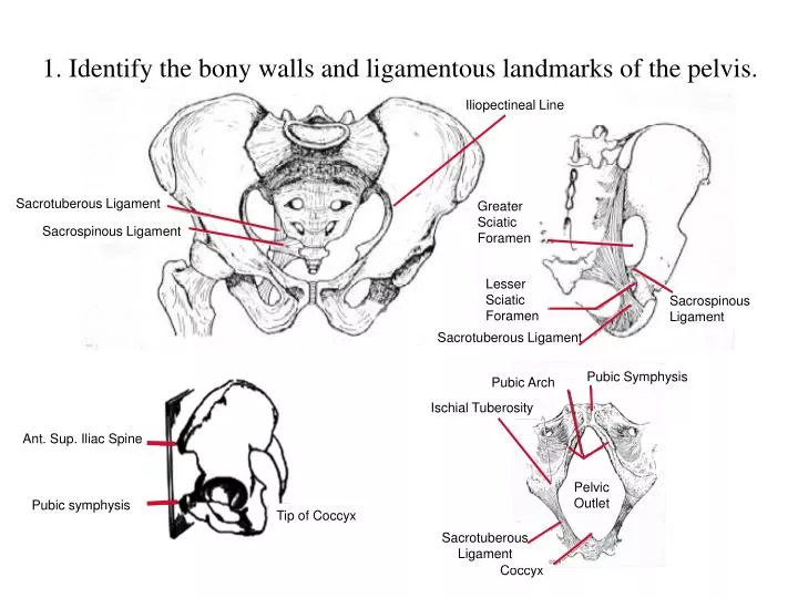
Iliopectineal Line. Pubic Symphysis. Pubic Arch. Ischial Tuberosity. Ant. Sup. Iliac Spine. Pelvic Outlet. Pubic symphysis. Tip of Coccyx. Sacrotuberous Ligament. 1. Identify the bony walls and ligamentous landmarks of the pelvis. Sacrotuberous Ligament. Greater Sciatic Foramen.

E N D
Iliopectineal Line Pubic Symphysis Pubic Arch Ischial Tuberosity Ant. Sup. Iliac Spine Pelvic Outlet Pubic symphysis Tip of Coccyx Sacrotuberous Ligament 1. Identify the bony walls and ligamentous landmarks of the pelvis. Sacrotuberous Ligament Greater Sciatic Foramen Sacrospinous Ligament Lesser Sciatic Foramen Sacrospinous Ligament Sacrotuberous Ligament Coccyx
Female Male Pelvic Inlet Pelvic Outlet Pelvic Cavity Pelvic Arch Male vs. Female Pelvis
Greater Sciatic Foramen Piriformis Muscle Piriformis Muscle Sacrospinous Ligament Sacrotuberous Ligament Sacrotuberous Ligament Gemellus Superior & Inferior Greater Trochanter Ischial Tuberosity Lesser Trochanter Muscular Walls Obturator Internus
4. Identify the pelvic diaphragm and its components Pubic Symphysis Levator Prostatae or Sphincter Vaginae Sphincter Vaginae (or Levator Prostatae) Urethra Pubic Symphysis Vagina Puborectalis Rectum Pubococcygeus Puborectalis Perineal Body Pubococcygeus Obturator Internus Coccyx Iliococcygeus Coccygeus Iliococcygeus Coccygeus Coccyx Sacrum Superior View Inferior View
Anteverted Anteflexed 2. Identify the normal position and anatomical relationships of the pelvic viscera
3. Identify the extent of the peritoneum and its folds and reflections in the male and female pelvis and their relationship to the pelvic contents. Rectovesicle pouch Vesicouterine pouch Rectouterine pouch Infraperitoneal space • Most pelvic organs are infraperitoneal
Peritoneal ligaments Pelvic visceral ligaments Fundus Bladder Bladder Lig. Of ovary Pubocervical lig. Round lig. of uterus Uterine tube Transverse cervical lig. Broad ligament Cervix Suspensory lig. Sacrocervical lig. Rectum Sacrocervical lig. Ovarian art. Uterine tube Lig. Of the ovary Mesosalpinx Bladder Lig. Of the ovary Fundus Round lig. of the uterus Rectum Round ligament of the uterus Mesometrium Body Pelvic diaphragm Ureter Uterine art. Transverse cervical lig. Pubocervical lig. Uterine art. Cervix Ligaments supporting pelvic organs
Celiac Superior mesenteric Renal Inferior mesenteric Ovarian Superior rectal Common iliac Testicular Ext. iliac Int. iliac Inguinal ligament Median Sacral Femoral 5. Follow the flow of blood into an out of the structures of the pelvis and perineum. Rules: 1. All pelvic organs are supplied by branches of the internal iliac artery except the ovaries and the upper third of the rectum. 2. Venous drainage follows the arterial supply, including the portal tributary, the inferior mesenteric vein. 3. Portal caval anastomses are found at the inferior rectal veins.
Para-aortic External iliac Superficial Inguinal Internal iliac 6. Identify the lymphatic drainage of structures of the pelvis and perienum. • Rules: • Lymphatics drain toward lymph nodes along internal iliac veins, except for the ovary (para-aortic nodes), and superior portion of the rectum (inferior mesenteric nodes) • Perineum drains to superficial inguinal nodes
Ureter Ductus deferens Inguinal canal Bladder Ejaculatory duct Ampulla Seminal vesicle Prostate Bulbourethral gland Urethra Ductus deferens Epidiymis Testis 7. Follow the course taken by an ovum through the female reproductive tract and the pathway taken by a spermatozoon through the male reproductive tract. Fimbria Uterine tube Fundus Ovary Uterine cavity Body Cervix Fornix Vagina