Download
1 / 24
420 likes | 2.35k Views
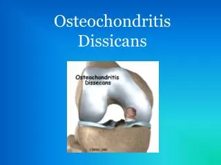
Osteochondritis Dissecans of the Ankle. November 24, 2003 Dr. N.C. Stone. Introduction. Incidence and Presentation Etiology and Mechanism of Injury Pathoanatomy Classification Imaging Treatment and Results. Incidence and Presentation. Osteochondral fracture of the Talar Dome

E N D
Osteochondritis Dissecans of the Ankle November 24, 2003 Dr. N.C. Stone
Introduction • Incidence and Presentation • Etiology and Mechanism of Injury • Pathoanatomy • Classification • Imaging • Treatment and Results
Incidence and Presentation • Osteochondral fracture of the Talar Dome • Males avg 25 years • Presents with ankle sprain • Initially missed (75%) • 2-6% of all ankle sprains
Incidence and Presentation • “ankle sprain not improving” • Stiffness, pain, effusion • Localized tenderness • Locking if loose fragment
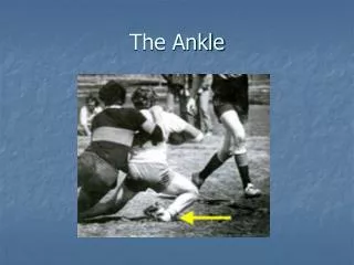
Etiology and Mechanism of Injury • Inversion injury • Lateral lesion, dorsiflexion, impacts and shears against fibula • Medial lesion, plantar flexed, posterior tibial plafond
Pathoanatomy • Once thought to be non-traumatic/AVN • Most agree now is traumatic • Osteochondral fragment is disrupted • If stable, new capillaries my cross fracture and revascularize fragment • If not stable or displaced, AVN and fragmentation
Classification • Burndt and Hardy (1959) • Many new MRI classifications
Imaging • 70% seen on plain films • Bone scan, CT are all used but MRI superior • Assess cartilegde, stability
Treatment • Stage 1 - rest, cast, non-operative • Stage 2 – same for 6 weeks, 90% good results • Stage 3 - lateral, definitely surgical – medial, more conservative • Stage 4 - surgical
Surgical Treatment • Acute 3 or 4 should have an attempt at repair • Peg, countersunk screws • Necrotic, fragmented, or small fragments • Excision, drill base
Surgical Treatment • Non-responding 1 and 2 • Drill but attempt to preserve articular surface
Surgical Treatment • Most can be done arthroscopically • +/- traction • Medial malleolar osteotomy may be necessary
OCD Long term • 88% good – excellent early • Best if < 1 year between injury and treatment • Lower grades do best
OCD Long term • Do poorly over time • Jensen et al. • After 9 years • 60% of patients had pain and stiffess • 90% mild arthrosis on radiographs
Conclusions • Ankle sprain that does not get better • MRI best • Non-surgical then surgical • Arthroscopically • Great early then poor long term prognosis