Download
1 / 3
30 likes | 104 Views
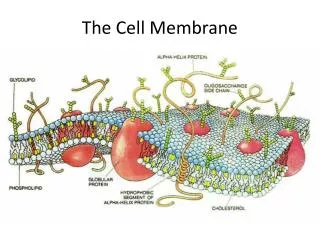
Microparticle 3D transportation on the membrane. The 1- m m bead is coated with fibronectin , which links to actin filaments through integrins .

E N D
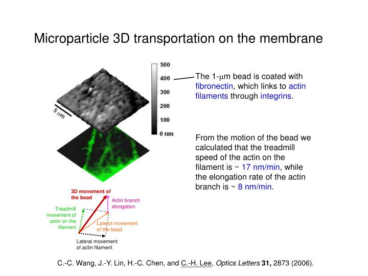
Microparticle 3D transportation on the membrane The 1-mm bead is coated with fibronectin, which links to actin filaments through integrins. From the motion of the bead we calculated that the treadmill speed of the actin on the filament is ~ 17 nm/min, while the elongation rate of the actin branch is ~ 8 nm/min. 3D movement of the bead Actin branch elongation Treadmill movement of actin on the filament Lateral movement of the bead Lateral movement of actin filament C.-C. Wang, J.-Y. Lin, H.-C. Chen, and C.-H. Lee, Optics Letters31, 2873 (2006).
600 400 200 m 10 mm 0 nm A gold nanoparticle coated by bovine serum albumin on the membrane NIWOP topography The nanoparticle moved into the membrane by ~ 400 nm, and then was expelled out, which indicates that the endocytosis of this particle did not complete. GFP-actin image Actin aggregates around the binding site of the gold particle (dia. = 80 nm). C.-C. Wang, J.-Y. Lin, C.-W. Lee, C.-Y. Huang, P.-K. Wei, and C.-H. Lee, “Observation of nanoparticle internalization on cellular membranes by using non-interferometric widefield optical profilometry,” submitted.
Observation of cancer cell filopodia without staining NIWOP bright field Bright field filopodium 2 mm 10 mm lung cancer cell Bright field + Restoration NIWOP + Restoration confocal fluorescence Filopodia are on the bottom of the cell. Lateral resolution is improved by using a maximum likelihood estimation algorithm to 130 nm. The filopodia can be observed clearly. T.-H. Hsu, W.-Y. Liao, P.-C. Yang, C.-C. Wang, J.-L. Xiao, and C.-H. Lee, Optics Express15, 76 (2007).