Download
1 / 67
680 likes | 1.09k Views
Non specific disease of parotid. Babak Saedi.MD Tehran university of Medical sciences Imam Khomeini Hospital. Anatomy & Physiology. Parotid Serous Sublingual Mucous Submandibular Mixed Minor salivary glands Controlled by sympathetic & parasympathetic . Acute & Chronic

E N D
Non specific disease of parotid Babak Saedi.MD Tehran university of Medical sciences Imam Khomeini Hospital
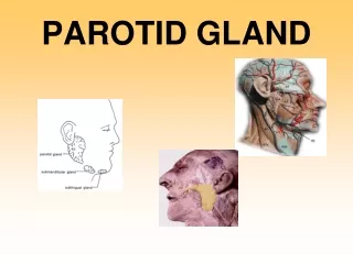
Anatomy & Physiology • Parotid • Serous • Sublingual • Mucous • Submandibular • Mixed • Minor salivary glands • Controlled by sympathetic & parasympathetic
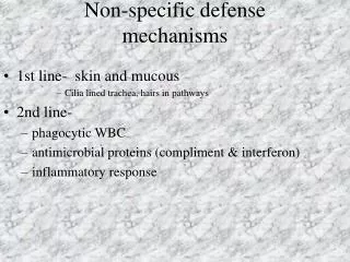
Acute & Chronic Non-Autoimmune Autoimmune Sialadenitis Necrotizing Sialametaplasia Sialadenosis Salivary Lymphoepithelial Cysts SALIVARY GLAND LESSIONS Non-Neoplastic Disease Benign Tumors Malignant Tumors Pleomorphic Adenomas Adenoid Cystic Carcinoma Basal Cell Adenomas Mucoepidermoid Carcinoma Myoepitheliomas Warthin’s Tumor Oncocytoma and Oncocytosis Sclerosing Polycystic Adenosis
Sialadenosis • Non-specific term used to describe a non-inflammatory non-neoplastic enlargement of a salivary gland, usually the parotid. • May be called sialosis • The enlargement is generally asymptomatic • Mechanism is unknown in many cases.
Sialadenosis (Sialosis) • Parotid glands most commonly. • Probably due to abnormalities of neurosecretory control.
Sialadenosis (Sialosis) • Cause maybe due to: • Nutritional (Alcoholism, Cirrhosis, Kwashiorkor and Pellagra • Endocrine (Diabetes, Thyroid diasease, Gonadal dysfunction) • Neurochemical (Vegetative state, Lead, Mercury, Iodine, Thiouracil)
Related to… • Metabolic “endocrine sialendosis” • Nutritional “nutritional mumps” • Obesity: secondary to fatty hypertrophy • Malnutrition: acinar hypertrhophy • Any condition that interferes with the absorption of nutrients (celiac dz, uremia, chronic pancreatitis, etc)
Related to… • Alcoholic cirrhosis: likely based on protein deficiency & resultant acinar hypertrophy • Drug induced: iodine mumps e. HIV
Sialadenosis (Sialosis) Histopathology: • Hypertrophy of serous acinar cells to about twice their normal size. • Cytoplasm is densely packed with secretory granules.
Allergic sialadenitis • Caused by drugs or allergens • Clinical presentation: • Acute salivary gland enlargement • Itching over the gland • With/without rash • Treatment • Self-limiting • Avoid allergen • hydration
ObstructiveSalivary Gland Disorders • Sialolithiasis • Mucous retention/extravasation
Mucocele9 • Mucus is the exclusive secretory product of the accessory minor salivary glands and the most prominent product of the sublingual gland. • The mechanism for mucus cavity development is extravasation or retention
Mucoceles & Ranula • Etiology • Trauma extravasation labial mucosa • Obstruction retention palate & floor of mouth • Clinical appearance • Ranula • extravasation / retention in floor of mouth • Obstruction of Sublingual salivary gland duct • Usually unilateral
Mucocele • Mucoceles, exclusive of the irritation fibroma, are most common of the benign soft tissue masses in the oral cavity. • Muco: mucus , coele: cavity. When in the oral floor, they are called ranula.
Mucocele9 Extravasation is the leakage of fluid from the ducts or acini into the surrounding tissue. Extra: outside, vasa: vessel Retention: narrowed ductal opening that cannot adequately accommodate the exit of saliva produced, leading to ductal dilation and surface swelling. Less common phenomenon
Mucocele • Consist of a circumscribed cavity in the connective tissue and submucosa producing an obvious elevation in the mucosa
Mucocele • The majority of the mucoceles result from an extravasation of fluid into the surrounding tissue after traumatic break in the continuity of their ducts. • Lacks a true epithelial lining.
Ranula9 • Is a term used for mucoceles that occur in the floor of the mouth. • The name is derived form the word rana, because the swelling may resemble the translucent underbelly of the frog.
Ranula9 • Although the source is usually the sublingual gland, • may also arise from the submandibular duct • or possibly the minor salivary glands in the floor of the mouth.
Ranula • Presents as a blue dome shaped swelling in the floor of mouth (FOM). • They tend to be larger than mucoceles & can fill the FOM & elevate tongue. • Located lateral to the midline, helping to distinguish it from a midline dermoid cyst.
Plunging or Cervical Ranula • Occurs when spilled mucin dissects through the mylohyoid muscle and produces swelling in the neck. • Concomitant FOM swelling may or may not be visible.
Treatment of Mucoceles9in Lip or Buccal mucosa • Excision with strict removal of any projecting peripheral salivary glands • Avoid injury to other glands during primary wound closure
Ranula Treatment9 • Marsupialization has fallen into disfavor due to the excessive recurrence rate of 60-90% • Sublingual gland removal via intraoral approach
Immunologic Disease Sjögren’s Syndrome7 • Most common immunologic disorder associated with salivary gland disease. • Characterized by a lymphocyte-mediated destruction of the exocrine glands leading to xerostomia and keratoconjunctivitis sicca
Sjögren’s syndrome7 • 90% cases occur in women • Average age of onset is 50y • Classic monograph on thediease published in 1933 by Sjögren, a Swedish ophthalmologist
SJOGREN’s SYNDROME All the above conditions plus; Dry eyes Generalized arthritis
Primary SS - Clinical picture • Mostly parotid gland is affected • Persistent / intermittent gland enlargement • bilateral, non-tender, firm, and diffuse swelling • saliva and altered saliva composition • Check of any recent changes to the character of the glands (nodularity) • significantly increased risk of developing B-cell lymphoma • Keratoconjunctivitissicca
Secondary SS - Clinical picture • Dryness of the skin & pruritis • Dry and persistent cough • >50% have arthralgia with or without arthritis • Dysphagia, nausea, dyspepsia, and epigastric pain • Peripheral & cranial neuropathy
Sjögren syndrome - Diagnosis • Different diagnostic criteria • Objective measurement of decreased salivary & lacrimal gland function • +ve autoimmune serologies • Minor salivary gland biopsy • Lymphocytic infiltration • Silagoraphy is also useful
Sjögren’s Syndrome • Keratoconjuntivitissicca: diminished tear production caused by lymphocytic cell replacement of the lacrimal gland parenchyma. • Evaluate with Schirmer test. Two 5 x 35mm strips of red litmus paper placed in inferior fornix, left for 5 minutes. A positive finding is lacrimation of 5mm or less. Approximately 85% specific & sensitive
Sjögren’s Lip Biopsy15 • Biopsy of SG mainly used to aid in the diagnosis • Can also be helpful to confirm sarcoidosis
Sjögren’s Lip Biopsy15 • Single 1.5 to 2cm horizantal incision labial mucosa. • Not in midline, fewer glands there. • Include 5+ glands for identification • Glands assessed semi-quantitatively to determine the number of foci of lymphocytes per 4mm2/gland
Sjögren syndrome - Treatment • Symptomatic • Systemic cholinergic (Pilocarpine) • 5mg TID/QID (should not exceed 30mg/day) • Follow up
Sjögren’s Treatment15 • Avoid xerostomic meds if possible • Avoid alcohol, tobacco (accentuates xerostomia) • Sialogogue (eg:pilocarpine) use is limited by other cholinergic effects like bradycardia & lacrimation • Sugar free gum or diabetic confectionary • Salivary substitutes/sprays
MICKULICZ’s SYNDROME 1) Symmetrical enlargement of salivary glands 2) Enlargement of the lachrymal glands 3) Dry mouth
Radiation induced pathology • Permanent salivary damage caused by doses 50Gy • Radioactive iodine for thyroid cancer treatment has similar but less severe effect • Clinical presentation • Salivary gland dysfunction signs & symptoms • Osteonecrosis • Increased risk of tumors affecting radiated tissues
Management steps for patients with radiation-induced xerostomia
Radiation Injury7 • Low dose radiation (1000cGy) to a salivary gland causes an acute tender and painful swelling within 24hrs. • Serous cells are especially sensitive and exhibit marked degranulation and disruption.
Continued irradiation leads to complete destruction of the serous acini and subsequent atrophy of the gland7. • Similar to the thyroid, salivary neoplasm are increased in incidence after radiation exposure7.
Granulomatous Disease7 Primary Tuberculosis of the salivary glands: • Uncommon, usually unilateral, parotid most common affected • Believed to arise from spread of a focus of infection in tonsils • Secondary TB may also involve the salivary glands but tends to involve the SMG and is associated with active pulmonary TB.
6- Granulomatous conditions • Tuberculosis • Granulation tissue formation in salivary gland • Xerostomia • Salivary gland enlargement • Sarcoidosis • Granulomas (T lymphocytes) affecting several organs • Lungs • Skin • Eyes • Parotid glands • Severity and duration of disease varies • Mild improvement noticed with steroid therapy
Granulomatous conditions • Tuberculosis • Granulation tissue formation in salivary gland • Xerostomia • Salivary gland enlargement • Sarcoidosis • Granulomas (T lymphocytes) affecting several organs • Lungs • Skin • Eyes • Parotid glands • Severity and duration of disease varies • Mild improvement noticed with steroid therapy
Granulomatous Disease7 Sarcoidosis: a systemic disease characterized by noncaseating granulomas in multiple organ systems • Clinically, SG involvement in 6% cases • Heerfordts’s disease is a particular form of sarcoid characterized by uveitis, parotid enlargement and facial paralysis. Usually seen in 20-30’s. Facial paralysis transient.
Granulomatous Disease7 Cat Scratch Disease: • Does not involve the salivary glands directly, but involves the periparotid and submandibular triangle lymph nodes • May involve SG by contiguous spread. • Bacteria is Bartonella Henselae(G-R) • Also, toxoplasmosis and actinomycosis.
Cysts7 True cysts of the parotid account for 2-5% of all parotid lesions May be acquired or congenital Type 1 Branchial arch cysts are a duplication anomaly of the membranous external auditory canal (EAC) Type 2 cysts are a duplication anomaly of the membranous and cartilaginous EAC
Cysts Acquired cysts include: • Mucus extravasation vs. retention • Traumatic • Benign epithelial lesions • HIV • Association with tumors • Pleomorphic adenoma • Adenoid Cystic Carcinoma • Mucoepidermoid Carcinoma • Warthin’s Tumor
Other: Pneumoparotitis • In the absence of gas-producing bacterial parotitis, gas in the parotid duct or gland is assumed to be due to the reflux of pressurized air from the mouth into Stensen’s duct. • May occur with episodes of increased intrabuccal pressure • Glass blowers, trumpet players • Aka: pneumosialadenitis, wind parotitis, pneumatocele glandulae parotis
Pneumoparotitis8 • Crepitation, on palpation of the gland • Swelling may resolve in minutes to hours, in some cases, days. • US and CT show air in the duct and gland • Consider antibiotics to prevent superimposed infection