Download
1 / 64
891 likes | 3.18k Views
Bacterial Anatomy. Rashmi.S. Anatomy of a Bacterial Cell. Description. Bacteria are Prokaryotic, unicellular that do not contain chlorophyll. Size of bacteria may range from 0.2-1.5 micrometer in diameter and 3-5 micrometer in length. DIFFERENCES BETWEEN PROKARYOTES AND EUKARYOTES.

E N D
Bacterial Anatomy Rashmi.S
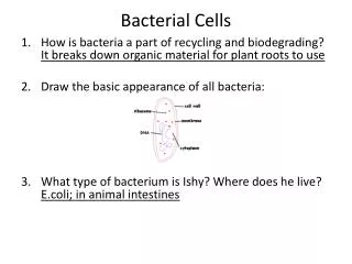
Description • Bacteria are Prokaryotic, unicellular that do not contain chlorophyll. • Size of bacteria may range from 0.2-1.5 micrometer in diameter and 3-5 micrometer in length
DIFFERENCES BETWEEN PROKARYOTES AND EUKARYOTES Character Prokaryotes Eukaryotes Nucleus: Nuclear Membrane Absent Present Nucleolus Absent Present Mitotic Division Absent Present Cytoplasm Cytoplasmic Streaming Absent Present Pinocytosis Absent Present Lysosomes Absent Present Golgi Apparatus Absent Present Endoplasmic Reticulum Absent Present Chemical Composition Sterols Absent Present Muramic Acid Present Absent Teichoic Acid Present Absent
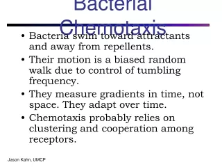
Classification of Bacteria based on their Shape • Cocci • Bacilli • Vibrio • Spirilla • Spirochetes • Actinomycetes • Mycoplasma
Cellular Arrangement In Cocci, • Diplococci: Cocci arranged in pairs • Streptococci: Arranged in chains • Staphylococci: Arranged in grape like clusters
In Bacilli, • Coccobacilli: Oval shaped • Palisades : Parallel, attached at any one end of the cell • Streptobacilli: In chains
Vibrio Spirilla
Mycoplasma Actinomycetes
Structure of a Bacterial Cell(Bacterial Anatomy) • Examination of a bacterial cell reveals components of structures • Some external to cell wall • Others internal to cell wall
Demonstration of the cell wall • Plasmolysis • Microdissection • Specific Antibodies • Differential Staining • Electron Microscope
Structure of the cell wall • Bacterial cell wall provides structural integrity to the cell. • The bacterial cell wall differs from that of all other organisms by the presence of Peptidoglycan • Peptidoglycan (Mucopeptide) is composed of alternating chains of .. • N -Acetyl Glucoseamine and N-Acetyl Muramic acid, which is cross linked by Peptide chains
Peptidoglycan is responsible for the rigidity of the bacterial cell wall and for the determination of cell shape • Based on the composition of cell wall & Staining bacteria are classified into “Gram positive” and “Gram Negative’
Gram Positive Cell wall • The Gram positive cell wall is characterized by the presence of a very thick Peptidoglycan layer • 20-80 nm thick • Cell wall contains90% Peptidoglycan and 10%Teichoic acid
Interwoven in the cell wall of gram-positive are Teichoic acids and lipoteichoic acids. • Teichoic acids composed of polymers of glycerol, phosphates, and the sugar alcohol- ribitol.
Teichoic acids constitute for the major surface antigens. Eg: In Streptococcus pneumoniaeTeichoic acid bears the antigenic determinants called the “Forssman antigen”
Gram Negative Cell Wall • Gram negative cell wall contains a thin Peptidoglycan layer adjacent to the Cytoplasmic membrane, • In addition to the Peptidoglycan layer, the Gram negative cell wall also contains an additional outer membrane composed by Phospholipids and Lipopolysaccharide which face into the external environment.
The LPS present on the Gm negative cell wall consists of 3 regions: • Polysaccharide determining O antigen • Core Polysaccharide • Glycolipid portion /Lipid A
All the three factors are responsible for the endotoxic activities……… • LPS Endotoxin causes a form of Septic Shock for which there is no direct treatment
Cytoplasmic Membrane • It is a thin layer lining the inner surface of the cell wall. • Semipermiable membrane controlling the flow of metabolites • Chemically ,consists of Lipoprotein and carbohydrates. Sterols are absent
Cytoplasm • Colloidal system of variety of organic and inorganic solutes in Viscous watery solution • No ER,& Mitochondria • Contains Mesosomes Inclusions and Vacuoles
Mesosomes • Vesicular, convoluted invaginations of the plasma membrane • Prominent in GM+ bacteria • Principal sites of Respiratory enzymes • Analogous to mitochondria in Eukaryotes
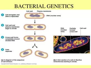
Nucleus • Bacterial nuclei “have no nuclear membrane “or the nucleolus. • Genome consists of a single double stranded DNA. • Might be a Circular form or may be open under certain condition to form a long chain.
Plasmids • Extra chromosomal DNA • Circular capable of autonomous replication. • Transferred from one bacterium to another .
Importance • Their presence confers certain special characters…… • Toxigenicity • Antibiotic Resistance • Ability to use certain unusual components as nutrients
Structures external to Bacteria • Capsule • Flagella • Pili(Fimbriae)
Capsule • Viscid material secreted by bacteria around the cell surface • Capsule is a sharply defined, organized structure (Eg: Pneumococcus) • Loose undemarkated structure as in Lueconostoc is a Slime layer.
Most bacterial capsules are composed of Polysaccharides Eg: Klebsiella pneumoniae • A few capsules are Polypeptides Eg: Bacillus anthracis
Quellung Reaction • Described by Neufeld(1902). • Serological method of demonstrating the capsule. • Suspension of capsulated bacterium is mixed with its specific anticapsular serum & examined under the microscope ,capsule appears prominent & swollen. • Used to type Pneumococci.
Functions of Capsules • Antiphagocytic,thus contribute “Virulence”. • Protects against “Lysozyme” • Promote attachment of bacteria to surface(Eg: Streptococcusmutans). • Permits bacteria to adhere to Medical Implants & Catheters.
Toxicity to host cell – Eg: Bacteroidesfragilis. • Provide protection against temporary drying. • Block the attachment ofBacteriophages.
Applications: • Used in serological typing • Detection of capsule in Blood, CSF provides a rapid method of diagnosis • Used in preparation of vaccines Eg: H.influenzae
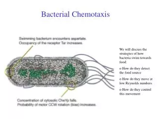
Flagella • Unbranched, long ,filaments ,made up of protein “Flagellin” • Organs of locomotion • Found in all motile bacteria except Spirochetes
Flagella are highly antigenic, • Termed as the ‘H’ Antigen. • Some of the immune responses are directed against these proteins.
Structure • 3-20 Micrometer. • Each flagellum consists of 3 parts 1.Filament 2.Hook 3.Basal body
Kinds of Motility: • Darting motility : V.cholerae • Tumbling motility: L.monocyctogenes • Cork &screw motility: T.pallidum • Stately motile : Clostridium spp. • Serpentine motility: Salmonella (Except S.gallonarum pullorum)