Download
1 / 48
550 likes | 1.11k Views
Dementia. Pratik Bhattacharya MD, MPH Chief of Neurology and Director: Stroke Program, Sinai-Grace Hospital. Assistant Professor, Department of Neurology, Wayne State University. Defining concepts: Memory. Explicit memory: requires conscious recall and fast acquisition.

E N D
Dementia Pratik Bhattacharya MD, MPH Chief of Neurology and Director: Stroke Program, Sinai-Grace Hospital. Assistant Professor, Department of Neurology, Wayne State University
Defining concepts: Memory • Explicit memory: requires conscious recall and fast acquisition. • Semantic memory: facts, objects and abstract concepts (e.g. geography) that have to be learned • Episodic memory: knowledge of past events • Implicit memory: does not require conscious recall • Procedural memory (learned over time – brushing teeth) • Anterograde amnesia: knowledge for experiences that occur after a certain event; inability to acquire new information • Retrograde memory: previously acquired information. • Mainly hippocampus, limbic system
Defining concepts: Attention • Attention networks different from memory networks • Visual, auditory stimuli • Drives your attention to it • Anatomy: • Inferior posterior parietal cortex (primary and association sensory cortex) • Prefrontal cortex (frontal eye fields) • Caudate nucleus
Defining Dementia Confusional State: Inability to think with proper speed and clarity, impaired immediate recall, decreased attention and concentration. Delirium: An acute confusional state with prominent alterations of perception and consciousness with hallucinations, delusions, heightened alertness and agitation, hyperactivity of psychomotor and autonomic functions, insomnia etc. Dementia: A syndrome characterized by deterioration of memoryplus two cognitive domains (executive function, praxis, language et cetera) compared to baseline; severe enough to interfere with usual social functioning and activities of daily life
Delirium • Usually altered sensorium, confusion, inattention, disorientation of acute/ subacute onset. • If chronic: generally called encephalopathy • Fluctuations over minutes or hours • Attention deficits: • Decreased ability to focus, sustain or shift attention • Distractible: unable to maintain attention to a particular task, but more attentive to trivial stimuli instead
Delirium: other key features • Disorganized thinking, disorganized content of speech with poor comprehension. • Hallucinations: often visual • Delusions: often persecutory; agitated patient may need restraints. • Altered sleep-wake cycle: reversal/ excessive daytime drowsiness. Sundowning. • Psychomotor hypoactivity (lethargy) fluctuating with psychomotor hyperactivity (agitation)
Alzheimer’s DiseaseRisk factors • Age • Female sex (survival effect) • Low level of education • Head trauma: repeated or severe. • Dementia pugilistica • Down’s syndrome • Patients with Down’s syndrome eventually develop AD • Mothers of children with Down’s syndrome are at greater risk for AD than general population
Alzheimer’s DiseaseRisk factors • APO ε4 susceptibility gene • Homozygous: increased risk for AD by age 85 • Effect is primarily on age of onset of symptoms • Seen in heterozygotes also • Many people with APO ε4 never develop the disease; so should not be used for diagnosis or risk assessment (AAN recommendation)
Alzheimer’s DiseaseRisk factors • Genetic and Familial disposition • Family history of dementia in first degree relatives • Autosomal dominant (present 40-70 years) • Familial AD susceptibility genes: • Chromosome 21 (Amyloid Precursor Protein) • Chromosome 14 (presenilin 1) • Chromosome 1 (presenilin 2) • Other candidate genes: insulin degrading enzyme (IDE)
Alzheimer’s Disease: Clinical Features • Early memory deficit • Impaired episodic memory, reduced delayed recall • Early stages called “amnestic Mild cognitive impairment” • Language deficits • Anomia, poor comprehension, semantic paraphasias • Visuospatial impairment: difficult visual processing • Balint’s syndrome: Oculomotorapraxia, optic ataxia, simultagnosia • Apraxia • Agnosia: mainly visual • Impaired executive function • Difficulty in performing activities of daily living
Alzheimer’s Disease: Clinical Features- Psychiatric symptoms • Early on: social withdrawal • Indifference and apathy • Depression – • Alzheimer requires pharm more than therapy • AD patients at higher risk due to loss of noradrenergic neurons in locus ceruleus. • Hallucinations: visual (more common w/ Alzheimers)or auditory • Phantom Boarder: • Theft of property; intruder living in the house • Capgras’ syndrome • Caretaker or family member is an imposter • Fregoli syndrome • Strangers are identified as family members
Alzheimer’s disease:Pathology: Atrophy • Neuronal loss: • First in the hippocampus and entorhinal cortex • Early involvement of Layers II and IV of CA1 and subiculum • Later: parietal neocortex, frontal, temporal, limbic cortex
Alzheimer’s disease:Pathology: Neurofibrillary Tangles • Tangle stages correlate with clinical picture • Paired helical filaments of hyperphosphorylated tau • First appears in Medial temporal structures • Ghost tangle: aggregates of tau eventually cause neuronal death: tangle remains behind. • Note: many other diseases have tau aggregates : Tauopathies.
Alzheimer’s disease:Pathology: Neuritic Plaques • Does not correlate with clinical severity • Extracellular • Loose aggregates of amyloid and preamyloid material • Mature plaque: focal aggregate: dense core of amyloid with surrounding halo of dystrophic tau positive neurites
Alzheimer’s disease:Pathology: other features • Granulovacuolar degeneration • Vacuoles inside neurons • Mainly hippocampal pyramidal cells • Also seen with Down’s syndrome and aging • AmyloidAngiopathy • Amyloid deposition in wall of small and medium sized vessels and leptomeningeal arteries • increased risk of hemorrhage
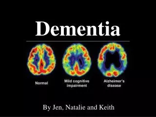
Alzheimer’s disease: Imaging-MRI Generalized atrophy Medial temporal lobe atrophy / hippocampal atrophy
Alzheimer’s disease: Imaging- Amyloid imaging • Pittsburgh Compound B • Stains amyloid • Widely used in AD research • Florbetapir • Binds to amyloid plaques • Identifies patients even before they have cognitive problems
Alzheimer’s Disease:Treatments- mild to moderate dementia Cholinesterase inhibition (Acetylcholinesterase) • Donepezil (Aricept) • Once daily regimen 5 mg/d for 1 month, then 10 mg/day • Nausea, vomiting, diarrhea, vivid dreams • Galantamine (Reminyl) • 4mg twice a day; raise up to 12 mg twice a day • More nausea, vomiting, vivid dreams • Rivastigmine (Exelon) • Also inhibits butyrylcholinesterase and ACh • 1.5 mg twice a day and raise up to 6 mg twice a day • Highest side effect profile; therefore patches available to reduce nausea and vomiting
Alzheimer’s Disease:Treatments- moderate to severe dementia • Memantine • NMDA antagonist • Activation of NMDA receptor improves memory and learning • Can be used as monotherapy or in combination with cholinesterase inhibitors and Vitamin E • Other symptomatic treatment of depression, psychosis, agitation. • Plenty of investigational therapies: immunization
Fronto-Temporal dementias (FTD)Clinical features • Men = Women • Disease duration 8 years (2-15) • Behavioral/ psychiatric • Personality change • Disinhibition • Antisocial • Emotional blunting • Mental rigidity • Executive dysfunction • Perseveration/ stereotypic actions • Decline in hygiene – Don’t car • Preservation of memory and visuospatial function till later
Fronto-Temporal dementias (FTD)Clinical features • KluverBucy syndrome • Bilateral anterior temporal lobe and amygdala • Hyperorality – Like to eat a lot… anything • Exploratory oral behavior • Weight gain • Hypo or hypersexuality • Hypermetamorphosis: touching and examining minor environmental stimuli.
Fronto-Temporal dementias (FTD)Clinical features: Progressive Non-Fluent Aphasia • Decreased speech output to mutism • Lack of grammar • Anomia • Phonemic paraphasia • Poor fluency with effortful and hesitant speech • Oral apraxia and stuttering • Associated alexia and agraphia • MRI: left fronto temporal atrophy.
Fronto-Temporal dementias (FTD)Clinical features: Genetic phenotypes • 20% are inherited. • Autosomal dominant • Mutations in the Tau gene on chromosome 17. • FTD 17 • Associated with parkinsonism. 4R Tau • FTD-MND • Associated with motor neuron disease. Chromosome 9 • FTD-ldh • Lacking distinctive histopathology: “ubiquitin positive, tau negative inclusions” • FTD-Picks disease • Pick’s inclusions: predominantly 3R Tau. No known mutation in the Tau gene. • FTD/AD • Features of AD also. Probably related to the APO E4 allele
Fronto temporal dementiasTreatment approaches • Symptomatic treatment of behavioral symptoms • SSRI • Antipsychotics: risperidone, olanzapine, quetiapine. • Inhibitors of Tau phosphorylation (experimental) • Lithium • Valproic Acid
Dementia with Lewy BodiesPathology • Alpha synucleindeposits. • CA1 and CA2 of hippocampus • Also in substantianigra, limbic regions, brainstem and neocortex • Fewer senile plaques and neurofibrillary tangles than AD • Eosinophilic neuronal inclusion with a halo.
Dementia with Lewy BodiesImaging: anatomic basis • MRI • Whole brain atrophy • Less than would expect with AD • PET • Hypometabolism especially in occipital lobe • Less extent in temporoparietal lobe.
Dementia with Lewy BodiesClinical Features • Progressive cognitive decline • Memory loss prominent but not in early stages • Fluctuations in attention and alertness • Recurrent visual hallucinations (well formed and detailed) • Motor symptoms of Parkinsonism • Neurolepticsensitivity • If you give Haldol for hallucinations will cause adverse reaction to anti-psychotic meds. Parkinsons worse • Use SGA – Second Generation Anti-psychotic instead • REM sleep behavior disorder. • Kick and punch in their sleep
Dementia with Lewy BodiesTreatment approach • Donepezil, galantamine, rivastigmine: effective for cognitive symptoms • Parkinsonism: Levodopa • Hallucinations: atypical neuroleptics: quetiapine, olanzepine, risperidone • SSRIs • Clonazepam and melatonin for REM sleep behavior disorder
Vascular DementiaClinical spectrum • Heterogenous group of disorders due to cerebrovascular disease. • Multiple ischemic infarcts • Single strategic infarct: • Caudate infarcts ( look like fronto-temporal dementia) • Hippocampus (memory difficulties) • Bithalamus (coma/ abulia)
Vascular DementiaClinical spectrum • Binswanger disease • Extensive diffuse white matter changes due to hypertension • Frontal executive dysfunction • Apathy and amotivational syndrome • CADASIL • Cerebral autosomal dominant arteriopathy with subacute infarcts and leuco-encephalopathy.
Vascular DementiaTreatment strategy • Antihypertensive disease • Calcium channel blockers may have a neuroprotective effect • Anticholinesterase inhibition • Data may be clouded by concomitant AD symptoms • Depression is quite common: • SSRI
Prion DiseaseCreutzfeldt-Jakob Disease • Prions are free surviving proteins. • When they infect cells, they alter the structure and function of normal cell protein: PrP • PrPsc is the abnormal form, accumulates in neurons • 85% are sporadic CJD • 15% are autosomal dominant • Dementia, myoclonus, pyramidal and extrapyramidal signs. • Death within one year- VERY VERY FAST DIMENTIA
Creutzfeldt-Jakob DiseaseHistopathology: Spongiform encephalopathy
CreutzfeldtJakob diseaseInvestigations • CSF • 14-3-3 protein levels: marker of neuronal loss • Non specific • MRI • Cortical ribbon sign • Pulvinar sign • EEG • Periodic sharp waves every 0.5 to 1 second • Myoclonic jerks • Biopsy
Iatrogenic CJD • Contaminated corneal transplants • Contaminated pituitary hormones • Dural grafts from cadavers • Pericardial grafts • Inadequate autoclaving of surgical instruments (134 C steam autoclaving)
Normal Pressure Hydrocephalus(adult hydrocephalus syndrome) • Triad: Gait apraxia (magnetic gait), urinary incontinence and dementia (frontal executive dysfunction) • Chronic history • Not really normal pressure: on prolonged monitoring- intermittent transient elevations of CSF pressure.
Normal Pressure Hydrocephalus • MRI: • Large ventricles • Periventricular white matter changes: transependymal edema due to CSF
Normal Pressure Hydrocephalus • Large volume tap 20-50 cc • Relieve pressure by removing fluid • Examination 1 hour and 24 hours after tap • See if it improves, if not, they can shunt • Shunt procedures • Gait and incontinence tend to improve; • dementia less likely to improve.
Questions? • pdbhatta@med.wayne.edu