Download
1 / 19
190 likes | 209 Views
This study analyzes the transition zone between main vessel and side branch in human coronaries to reveal missed geometrical insights. Detailed geometric analysis was performed using casts of human coronary trees, showing asymmetry and unique features to guide stent design and improve outcomes. The study provides valuable data on bifurcation anatomy, shaping stent implantation strategies.

E N D
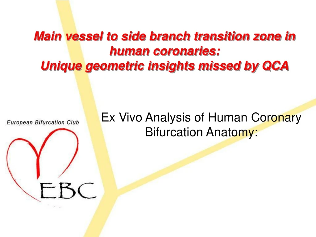
Main vessel to side branch transition zone in human coronaries: Unique geometric insights missed by QCA Ex Vivo Analysis of Human Coronary Bifurcation Anatomy:
Mary E. Russell, MD, FACC Ascent Translational Sciences Carlisle, Massachusetts, USA Eitan Konstantino, PhDGary Binyamin, PhD TriReme Medical, Inc Pleasanton, CA, USA
Technical Challenges with BifurcationsUsing Straight, Concentric Tubular Systems
Program Objectives Define bifurcation anatomy and geometry • Casts of human coronary tree to evaluate intersection between Main Vessel (MV) & Side Branch (SB) • Qualitative assessments • Shapes at the SB take off • Quantitative measures • Specified Diameters (vessels > 1.6 mm) • Various angles
Methods • Retrograde aortic polymer injection to create casts of the human coronary arteries • 23 Adult Cadaver Heart Donors: • 11 Females, 12 Males • Predominantly white • Mean age: 66 (range: 56-76) • Causes of death: noncardiac in 22 of 23 cases • 27 vessels with side branches >1.99 • 70 vessels with side branches >1.65 mm
Cast Preparation • Aorta perfused retrograde ≥ 60 min • Polymer injected to fill the aortic sinus and coronary arteries • Polymer cured (≥ 60 min) • Models immersed in caustic solutions to digest tissue
LAD LM LCX RCA 3 Dimensional Casts of Coronary Tree(Aorta to terminal branches (<1mm) • Branching • Curvature • Tortuosity • Lesions • Intersections
Geometric Analysis Performed by Imaging(MV to SB intersection) • Smartscope MVP100 video-based inspection system • Gage-X metrology software • Magnification ≥ 34 X (novel view between angiographic & microscopic) • Measurements made with bifurcationperpendicular to the field of view • Two independent reviewers performed measures if discordant by >10% then, consensus review
High Power Views of Anatomy & DiseaseMultifaceted intersection without discrete angle No disease Minor stenosis; minimal disease Moderate ostial stenosis; diffuse stenosis in SB and proximal MV Severe stenosis and disease
conical taper elliptical Ostial Geometry: Oval and Asymmetric Rather than Round Example: Side Branch of RCA Side view of ostium with SB removed Front view of ostium with SB removed Sketches of ostium
Side branch diameter Ostial diameter 3 mm 3 mm 3 mm Proximal diameter Distal diameter Center point Diameter Measurements Note: Illustration not to scale
Diameters: Greater proximal to distalOstial SB diameter similar to distal MV Main Vessel 1.70 - 4.18 mm Side Branch1.59 - 2.59 mm 2.86 2.39 2.29 1.93
Ostial Geometry:Transition Zone Taper Greater by 3-fold Average Taper Vessels with SB > 1.99 mm (Main Vessel) Proximal to Distal Taper (Side Branch) Ostium to Side Branch Taper Main Vessel Tapers 0.56 mm over 6.00 mm distance Side BranchTapers 0.60 mmover 1.75 mm distance
Proximal radius of curvature Proximal transition angle Distal radius of curvature Distal transition angle Proximal Intersection angle Distal Intersection angle Intersection Angles: intersection of centerline of main vessel and side branch (~measured by angiography) Transition Zone Angles: actual main vessel to side branch progression as measured from the main vessel Three Types of Angle Measurements Note: Illustration not to scale
Intersection Angles: Proximal Distal Intersection Angles*:Obtuse Proximal vs Acute Distal Proximal angles (77°-178°; mean 134°) Distal angles (12°-99°; mean 54°) *Intersection angles = angiographic measurements
Transition Angles: Proximal Distal Transition Zone Angles*:Differential smaller between proximal & distal angles Proximal transition angles (107°-177°; mean 152°) Distal transition angles (27°-163°; mean 109°) *Transition zone angles = actual main vessel to side branch progression as measured from the main vessel
Summary Bifurcation diameters (~ to previous reports) MV:Wide Range (1.7 to 4.2), proximal mean= 2.86 distal mean= 2.39 SB: Wide Range (1.6 to 2.6), mean 2.28 Fours Types of Asymmetric Ostial Geometry: • Multifaceted transition (high magnification detail) • Oval rather than round ostium • Taper with SB 3-fold greater than MB • Side branch take off angles • Proximal (obtuse) • Distal (acute)
ConclusionsDistort stent or Distort anatomy? • Complex transition zone from the MV to SB up to 4 asymmetric features • Anatomic distortion likely with symmetric (cylindrical) designs • Strut protrusion/injury • Gaps • Incomplete wall apposition • Matching design to asymmetric ostial geometry may minimize implant injury, enhance scaffolding and improve outcomes
Four types of asymmetryImplications—more questions Angiography (uniform definitions needed) • What, where and how angles measured • Usefulness of measures? IVUS of SB • Is SB imaging essential? • Is the oval shape seen at the SB take off really an artifact due to cathetor angulations on w/drawal? • How should strut apposition be measured in the MV to SB transition zone Procedural • Stent designs to match MV to SB transition zone? • Post dilation balloons & protocols that maintain elliptical sshpae &differential SB taper?