Download
1 / 39
710 likes | 2.43k Views
ANATOMY AND PHYSIOLOGY OF THE URINARY SYSTEM. OBJECTIVES. Identify the main functions of the urinary system Learn the organs of the urinary system and describe their physical characteristics Describe the microscopic structure of the kidney and learn the function of the nephron

E N D
OBJECTIVES • Identify the main functions of the urinary system • Learn the organs of the urinary system and describe their physical characteristics • Describe the microscopic structure of the kidney and learn the function of the nephron • Learn causes and symptoms of renal failure and describe different types of dialysis • List common diseases of the urinary system
INTRODUCTION • Urinary system works together with circulatory, digestive and endocrine systems to remove waste and to maintain homeostasis.
INTERESTING FACTS • Each kidney has over 1 000 000 nephrons and about 140 miles of filters and tubes. • Each minute kidneys filter over 1000 cc of blood • Each hour kidneys are producing about 60 cc of urine • Each day a person takes in approximately 2500 cc of fluids • Each day a person eliminates approximately 1500 cc of urine
FUNCTIONS OF THE URINARY SYSTEM • Excretion- removal of waste products from blood • Secretion - production and transportation of urine • Elimination – emptying urine from the urinary bladder
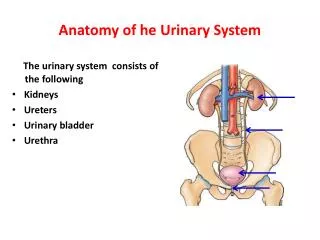
ORGANS OF THE URINARY SYSTEM • Kidneys • Ureters • Urinary bladder • Urethra
KIDNEYS • Two approximately 4.5 inches bean shaped organs • Located on either side of the vertebral column • Kidneys are positioned behind parietal peritoneum • A heavy cushion of fat protects and keeps kidney in their proper position • Outside they are covered by a tough fibrous capsule
MACROSCOPIC STRUCTURE • Concave medial side of the kidney is called hilus • The renal artery and nerves enter kidney here • The renal vein and ureter exit here
INTERNAL STRUCTURE • Cortex – outer layer • Medulla – inner layer • Pyramids – triangular divisions of the medulla • Pelvis – cavity within the kidney that receives the urine
MICROSCOPIC STRUCTURE NEPHRON – the microscopic structural and functional unit of the kidney There are about 1.25 million nephrons in each kidney
MICROSCOPIC STRUCTURE • Its chief function is to regulate the concentration of water and soluble substances like sodium salts by filtering the blood • Reabsorbing what is needed and excreting the rest as urine. • A nephron eliminates wastes from the body • Regulates blood volume and blood pressure • Controls levels of electrolytes and metabolites • Regulates blood pH
MICROSCOPIC STRUCTURE • Bowman’s capsule – cup shaped top of nephron • Glomerulus- network of about 50 blood capillaries • Renal tubules – receive and concentrate initial filtrate to produce final urine
URETERS • Two muscular tubes approximately 10-12 inches long • Function – to drain urine from the kidney to the bladder • Peristalsis moves urine in one direction only
URINARY BLADDER • Collapsible muscular sac, with folds in lining • Location- behind the pubic bones • Functions • Serves as reservoir for urine • Urge to void – when contains 250 ml • Max. capacity 1000 ml
URETHRA • Narrow tube leading from bladder to outside • External opening is called urethral meatus • Female urethra carries only urine and is 1.5–2 inches (4–5 cm) long • Male urethra carries both urine and semen, is about 8 inches (20 cm) long and opens at the end of the penis
TERMINOLOGY RELATED TO URINE AND URINATION • ANURIA – absence of urine production • RETENTION – inability to void urine • INCONTINENCE – inability to control urination • OLIGURIA – scanty urine output • POLYURIA – excessive urination • NOCTURIA – having to urinate at night • HESITANCY – difficulty in initiating urination • FREQUENCY – necessity to void often • URGENCY- sudden need to void
CONJENITAL ABNORMALITIES • Vesicoureteral reflux (VUR) is an abnormal movement of urine from the bladder into ureters or kidneys.
RENAL ECTOPIA • Renal ectopia - abnormal renal location
HORSESHOE KIDNEY • Intravenous urogram (IVU) demonstrates horseshoe kidney. • Note the malrotated collecting systems on both sides.
RENAL AGENESIS • Renal agenesis: DTPA scan shows agenesis of the right kidney
INFLAMMATORY DISEASES • Cystitis – inflammation of the urinary bladder • Causes – usually bacteria • Symptoms – frequency, dysuria, hematuria, abdominal pain, fever, lower back pain • Treatment – drink a lots of fluids, antibiotics • Urethritis – inflammation of the urethra • Ureteritis – inflammation of the ureters • Pyelitis – inflammation of the kidney’ pelvis
UTI – urinary tract infection • Cloudy or bloody urine, which may have a foul or strong odor • Low fever (not everyone will have a fever) • Dysuria • Pressure or cramping in the lower abdomen (usually middle) or back • Strong need to urinate often, even right after the bladder has been emptied • Flank (side), back, or groin pain • Flushed, warm, or reddened skin • Mental changes or confusion (in the elderly, these symptoms often are the only signs of a UTI) • Nausea and vomiting • Severe abdominal pain (sometimes)
UROLITHIASIS • A kidney stone is a solid mass made up of tiny crystals. • One or more stones can be in the kidney, ureter , bladder or urethra at the same time. • Causes: • Dehydration from reduced fluid intake • Strenuous exercise without adequate fluid replacement • Obstruction to the flow of urine • Kidney stones can also result from infection in the urinary tract • Gout results in chronically increased amount of uric acid in the blood and urine and can lead to the formation of uric acid stones. • Metabolic abnormalities • Some medications also raise the risk of kidney stones. These medications include some diuretics, calcium-containing antacids
UROLITHIASIS • Symptoms: • A kidney stone may or may not cause signs and symptoms until it has moved into the ureter • Severe pain in the side and back, below the ribs • Pain that spreads to the lower abdomen and groin • Pain on urination • Pink, red or brown urine - hematuria • Nausea and vomiting • Persistent urge to urinate • Fever and chills if an infection is present
DIAGNOSIS OF UROLITHIASIS • X rays with contrast medium • Ultrasound • Blood tests • Urine tests
TREATMENT • A simple and most important lifestyle change to prevent stones is to drink more liquids-water is best. Someone who tends to form stones should try to drink enough liquids throughout the day to produce at least 2 quarts of urine in every 24-hour period. • A doctor may prescribe certain medications to help prevent calcium and uric acid stones. • Extracorporeal Shock Wave LithotripsyIn ESWL, shock waves that are created outside the body travel through the skin and body tissues until they hit the denser stones. The stones break down into small particles and are easily passed through the urinary tract in the urine.
GLOMERULONEPHRITIS • Glomerulonephritis is inflammation of the filters in y kidneys (glomeruli). • Glomeruli remove excess fluid, electrolytes and waste from bloodstream. • Glomerulonephritis can be • acute — a sudden attack of inflammation • chronic — coming on gradually. • Symptoms: • Hematuria • Foamy urine due to excess protein (proteinuria) • High blood pressure (hypertension) • Fluid retention (edema) with swelling evident in your face, hands, feet and abdomen • Fatigue from anemia or kidney failure
TREATMENT • There is no specific treatment for the chronic form • Immunosupresive medications can be given
RENAL FAILURE • Renal failure or kidney failure describes a medical condition in which the kidneys fail to adequately filter toxins and waste products from the blood. • The two forms are: • acute • chronic • Symptoms: oliguria, severe electrolyte imbalance, anemia, uremia - condition resulting from kidney failure in which there is retention in the bloodstream of waste products normally removed by kidney
TREATMENT OF KIDNEY FAILURE • Dialysis is a process for removing waste and excess water from the blood. There are two types: • Hemodialysis • Peritoneal dialysis • Transplant