Download
1 / 24
240 likes | 368 Views
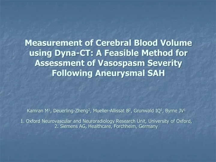
Kamran M 1 , Deuerling-Zheng 2 , Mueller- Allissat B 2 , Grunwald IQ 1 , Byrne JV 1 1. Oxford Neurovascular and Neuroradiology Research Unit, University of Oxford, 2. Siemens AG, Healthcare, Forchheim, Germany.

E N D
Kamran M1, Deuerling-Zheng2, Mueller-Allissat B2, Grunwald IQ1, Byrne JV1 1. Oxford Neurovascular and Neuroradiology Research Unit, University of Oxford, 2. Siemens AG, Healthcare, Forchheim, Germany Measurement of Cerebral Blood Volume using Dyna-CT: A Feasible Method for Assessment of Vasospasm Severity Following Aneurysmal SAH
Background • Image quality • Contrast resolution • Spatial resolution • Temporal resolution • Detector efficiency • Mechanical properties • of C-arm gantry Three dimensional volume Angiograms CT-like soft tissue images
Background • Image quality • Contrast resolution • Spatial resolution • Temporal resolution • Detector efficiency • Mechanical properties • of C-arm gantry Three dimensional volume Angiograms CT-like soft tissue images
Background Cerebral vasospasm following aneurysmal SAH An ideal technique for detection: Detect presence or absence of vasospasm before clinical deficits Be objective (reproducible) Effective in the unconscious patient Non-invasive
Background Cerebral vasospasm following aneurysmal SAH An ideal technique for detection: Detect presence of absence of vasospasm before clinical deficits Be objective (reproducible) Effective in the unconscious patient Non-invasive Diagnosis and assessment: Catheter angiography Transcranial Doppler Ultrasonography CT or MR perfusion scans
CBV measurement using C-arm FDCT Subtraction of the mask run from the contrast run Normalisation of parenchyma with an input function from major vessels CBV parametric maps Steady state 8 seconds Concentration Mask run Contrast run Time
C-arm FDCT Three dimensional volume
C-arm FDCT Three dimensional volume
Methods Dyna-CT • CBV Scan parameters: Two 8-sec acquisitions (mask and contrast enhanced), approx. 400 projections each at 0.5° steps, fluoroscopic monitoring in between • Post-processing for CBV estimation: co-registration, subtraction, normalisation with an input function • Angiographic reconstructions: contrast enhanced run only, 512×512 matrix, smooth/sharp kernel MR scan • MR-PWI, MR-DWI, Time of Flight MRA, T1, and T2 weighted sequences • Perfusion scan: DSC, T2* weighted gradient echo planar sequence (20ml 0.5M Gadolinium based contrast material), TR~2000msec, TE 44msec, FOV 248×248mm, 256×256 matrix, voxel size 0.98×0.98×4mm, 15 slices, 50 time-points • Perfusion analysis: block-circulant SVD algorithm, AIF chosen semi-automatically, motion correction
Example case 1 • A 60 year old lady presenting with ruptured anterior communicating artery aneurysm (5 × 7mm) • Day 0: SAH, no loss of consciousness, Grade 1 • Day 1: Coil embolisation, Grade 1 • Day 3: Fluctuating LOC, numbness of the left arm and leg, normal CT • Day 4: Weakness of both legs, neuro-ITU for supportive therapy MR-PWI and Dyna-CT scans Angioplasty
Example case 1 • A 60 year old lady presenting with ruptured anterior communicating artery aneurysm (5 × 7mm) • Day 0: SAH, no loss of consciousness, Grade 1 • Day 1: Coil embolisation, Grade 1 • Day 3: Fluctuating LOC, numbness of the left arm and leg, normal CT • Day 4: Weakness of both legs, neuro-ITU for supportive therapy MR-PWI and Dyna-CT scans Angioplasty MR-DWI MR-PWI MTT Dyna-CT CBV MR-PWI CBV
Example case 1 • A 60 year old lady presenting with ruptured anterior communicating artery aneurysm (5 × 7mm) • Day 0: SAH, no loss of consciousness, Grade 1 • Day 1: Coil embolisation, Grade 1 • Day 3: Fluctuating LOC, numbness of the left arm and leg, normal CT • Day 4: Weakness of both legs, neuro-ITU for supportive therapy MR-PWI and Dyna-CT scans Angioplasty MR-DWI MR-PWI MTT L R Dyna-CT CBV MR-PWI CBV
Example case 1 • A 60 year old lady presenting with ruptured anterior communicating artery aneurysm (5 × 7mm) • Day 0: SAH, no loss of consciousness, Grade 1 • Day 1: Coil embolisation, Grade 1 • Day 3: Fluctuating LOC, numbness of the left arm and leg, normal CT • Day 4: Weakness of both legs, neuro-ITU for supportive therapy MR-PWI and Dyna-CT scans Angioplasty MR-DWI MR-PWI MTT L R Dyna-CT CBV MR-PWI CBV
Example case 2 A 41 year old man presenting with ruptured anterior communicating artery aneurysm (6×4.5mm) • Day 0: SAH, Grade 1 • Day 6: Reached hospital. CT scan • Day 7: Dysphasia and right sided hemiplegia; neuro-ITU for supportive therapy. Deficits recovered. CTA showed an Acom (6x4.5x5 mm) and a left MCA (3x3.5x3 mm) aneurysm. • Day 7: Coil embolisation of both aneurysms. Vasospasm of ACA and MCA • Day 8: Deteriorating LOC, dysphasia, and weakness of right side of the body. CT, MR-PWI scan Dyna-CT CBV Angioplasty (Nimodipine)
Example case 2 A 41 year old man presenting with ruptured anterior communicating artery aneurysm (6×4.5mm) • Day 0: SAH, Grade 1 • Day 6: Reached hospital. CT scan • Day 7: Dysphasia and right sided hemiplegia; neuro-ITU for supportive therapy. Deficits recovered. CTA showed an Acom (6x4.5x5 mm) and a left MCA (3x3.5x3 mm) aneurysm. • Day 7: Coil embolisation of both aneurysms. Vasospasm of ACA and MCA • Day 8: Deteriorating LOC, dysphasia, and weakness of right side of the body CT, MR-PWI scan Dyna-CT CBV Angioplasty (Nimodipine)
Example case 2 A 41 year old man presenting with ruptured anterior communicating artery aneurysm (6×4.5mm) • Day 0: SAH, Grade 1 • Day 6: Reached hospital. CT scan • Day 7: Dysphasia and right sided hemiplegia; neuro-ITU for supportive therapy. Deficits recovered. CTA showed an Acom (6x4.5x5 mm) and a left MCA (3x3.5x3 mm) aneurysm. • Day 7: Coil embolisation of both aneurysms. Vasospasm of ACA and MCA • Day 8: Deteriorating LOC, dysphasia, and weakness of right side of the body CT, MR-PWI scan Dyna-CT CBV Angioplasty (Nimodipine)
Example case 2 R L A 41 year old man presenting with ruptured anterior communicating artery aneurysm (6×4.5mm) • Day 0: SAH, Grade 1 • Day 6: Reached hospital. CT scan • Day 7: Dysphasia and right sided hemiplegia; neuro-ITU for supportive therapy. Deficits recovered. CTA showed an Acom (6x4.5x5 mm) and a left MCA (3x3.5x3 mm) aneurysm. • Day 7: Coil embolisation of both aneurysms. Vasospasm of ACA and MCA • Day 8: Deteriorating LOC, dysphasia, and weakness of right side of the body CT, MR-PWI scan Dyna-CT CBV Angioplasty (Nimodipine)
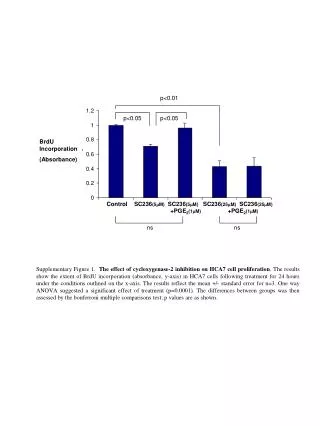
Results Grey matter r = 0.84 95% limits of agreement = -0.21 to +0.23 Measurement bias = 0.03 White matter r = 0.89 95% limits of agreement = -0.07 to +0.06 Measurement bias = -0.01
Results Grey matter r = 0.84 95% limits of agreement = -0.21 to +0.23 Measurement bias = 0.03 White matter r = 0.89 95% limits of agreement = -0.07 to +0.06 Measurement bias = -0.01
Results Overall (GM+WM) r = 0.87 95% limits of agreement = -0.16 to +0.17 Measurement bias = 0.02 Radiographic contrast: 80ml of Niopam 370 Radiation dose: 1.78 mSv Scan time: ~25 sec
Conclusion • Dyna-CT CBV agree with MR-PWI CBV • Improved spatial resolution, short scanning time, complete brain coverage • Exploits the same data to generate angiographic and soft tissue images (with reduced contrast and radiation dose) • Potentially useful tool that may help in time efficient triage of patients with brain ischaemia in optimized interventional environment However, • Improvements in detector efficiency and gantry rotation speeds are warranted to exploit its soft tissue and perfusion imaging potential
Acknowledgements • Department of Neuroradiology, West Wing, JR Hospital, Oxford • The Rhodes Trust • Siemens AG Healthcare, Forchheim Germany Many Thanks !