1 / 47

Shoulder Injuries by: Nanda K. Sinha, M.D.

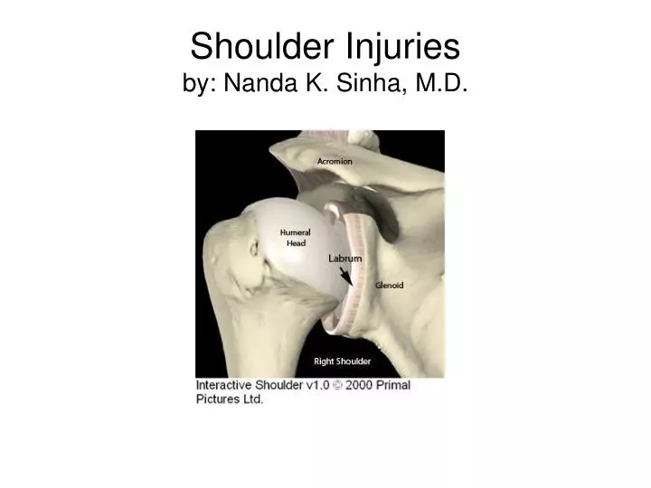
Shoulder Injuries by: Nanda K. Sinha, M.D. Surface Anatomy. Surface Anatomy. Surface Anatomy. Normal shoulder. Normal shoulder: axillary view. Normal xray-scapular Y view. Gleno-Humeral dislocation Anterior:Posterior. Gleno-Humeral dislocation Inferior.

mada
Download Presentation

Shoulder Injuries by: Nanda K. Sinha, M.D.

An Image/Link below is provided (as is) to download presentation Download Policy: Content on the Website is provided to you AS IS for your information and personal use and may not be sold / licensed / shared on other websites without getting consent from its author. Content is provided to you AS IS for your information and personal use only. Download presentation by click this link. While downloading, if for some reason you are not able to download a presentation, the publisher may have deleted the file from their server. During download, if you can't get a presentation, the file might be deleted by the publisher.

E N D

Presentation Transcript


  1. Shoulder Injuriesby: Nanda K. Sinha, M.D.

  2. Surface Anatomy

  3. Surface Anatomy

  4. Surface Anatomy

  5. Normal shoulder

  6. Normal shoulder: axillary view

  7. Normal xray-scapular Y view

  8. Gleno-Humeral dislocationAnterior:Posterior

  9. Gleno-Humeral dislocationInferior

  10. Gleno-Humeral dislocationY view of scapula

  11. Gleno-Humeral dislocation

  12. Gleno-Humeral dislocation

  13. Gleno-Humeral dislocation(shoulder dislocation) • Anterior: Most common • Posterior: Most missed • Inferior: Most dramatic • Superior: Acromion blocks superior displacement

  14. Gleno-Humeral dislocationReduction technique

  15. Gleno-Humeral dislocationReduction technique

  16. Gleno-Humeral dislocationReduction technique

  17. Gleno-Humeral dislocationReduction technique

  18. Minimally displaced fracture: non-operative management

  19. 4 part fracture

  20. 4 part fracture -Treatment

  21. displaced fracture: closed or open reduction: fixation

  22. Non-op vs operative

  23. Fracture: shaft of humerus

  24. Clavicle Fracture

  25. Clavicle Fracture • Medial ( inner) 1/3rd uncommon • Middle 1/3rd most common • Lateral (outer) 1/3rd

  26. Clavicle Fracturemiddle 1/3rd

  27. Clavicle FractureLateral 1/3rd

  28. Clavicle Fracture

  29. Clavicle Fracturecomminuted,displaced

  30. Clavicle Fracturex-ray 15 degree cranial

  31. Clavicle FractureIndications for ORIF • Shortening of 20 mm or more • Open injury • Impending skin disruption and irreducible fracture • Vascular compromise • Progressive neurologic loss • Displaced lateral 1/3rd fracture

  32. Clavicle Fracturearm sling vs fig of 8

  33. Clavicle Fractureopen reduction internal fixation (ORIF)

  34. Clavicle Fracturenon-op vs orif

  35. Clavicle Fracturenon op vs orif

  36. Clavicle Fracturenon-op vs orif

  37. Clavicle Fracturenon-op vs orif

  38. Acromio-Clavicular Dislocation(A-C Joint dislocation)

  39. Acromio-Clavicular Dislocation

  40. Acromio-Clavicular Dislocation

  41. Acromio-Clavicular Dislocation

  42. Acromio-Clavicular Dislocation

  43. Acromio-Clavicular Dislocation

  44. Acromio-Clavicular Dislocationtreatment • Grade 1 and 2: non operative • Grade 4 and above: ORIF • Grade 3: ( A-C Joint reducible by pushing up on the elbow at 90 degree) Non –op vs ORIF:

  45. Acromio-Clavicular DislocationNon-operative treatment • Sling • Ice • Pain medications • Activity limitation • Continue to monitor amount of displacement • Rehabilitation once pain is controlled

  46. Thank You!

More Related