Download
1 / 53
540 likes | 725 Views
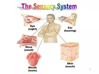
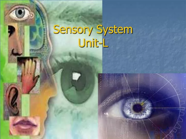
Sensory System Unit-L. Main Objectives. Describe the structure of the eye Analyze the function of the eye Describe the structure of the ear, nose, and tongue. Analyze the function of the ear, nose, and tongue. Analyze characteristics and treatment of common sensory disorders. The Eye.

E N D
Main Objectives • Describe the structure of the eye • Analyze the function of the eye • Describe the structure of the ear, nose, and tongue. • Analyze the function of the ear, nose, and tongue. • Analyze characteristics and treatment of common sensory disorders.
The Eye • 1” in diameter • Protected by orbital cavity of skull, eyebrows, eyelashes and eyelids. • Bathed in fluid from Lacrimal Glands ( tears empty into nasal cavity)
Cornea – Front of sclera • Clear structure called “window of the eye” (no blood vessels) • Transparent so light rays can pass through • Gets O2 and nutrients through lymph
A thin membrane that lines the eyelids and covers part of the eye. It secretes mucous to lubricate the eye. Conjunctiva
Wall of Eye is made up of 3 coats Sclera, Choroid Coat, and the Retina SCLERA- Outer layer White of the eye Tough, fibrous capsule helps maintain shape of eye and protects the structure within. .
Muscle responsible for moving the eye that are attached to the sclera. EXTRINSIC MUSCLES
Middle coat Contains blood vessels Circular opening in front is the PUPIL Colored, muscular layer surrounding pupil is IRIS Choroid Coat
Pupil constricts-gets smaller-in bright light Pupil dilates-gets larger-in dark light PUPILS
Theses muscles change size of iris & pupil to control amount of light entering the eye. The black center is a hole called the pupil. INTRINSIC MUSCLES
LENS • Crystalline structure located behind iris and pupil. • Elastic, disc-shaped, biconvex • Situated between the anterior and posterior chambers. • The part of the eye that is removed during cataract surgery. • Lies directly behind the pupil.
LENS • Accommodation is the function of the lens. It adjusts the focus of an eye to give clear vision
ANTERIOR CHAMBER filled with AQUEOUS HUMOR, a watery fluid. POSTERIOR CHAMBER filled with transparent, jellylike substance- VITREOUS HUMOR
Innermost layer Light rays focus an image on the retina The optic nerve is formed on the optic disc by nerve fibers. The image travels to the cerebral cortex via the OPTIC NERVE. Retina
Retina contains specialized cells- rods and cones. RODS- sensitive to dim light. CONES- sensitive to bright light and color. RODS & CONES
OPTIC DISC- on the retina, known as the blind spot- nerve fibers gather here to form the optic nerve, no rods or cones. Blind Spot
The Pathway of Vision CORNEA PUPIL LENS (Light rays are refracted) RETINA
The Ear Outer Ear • PINNA (AURICLE)- outer ear, collects sound waves • EXTERNAL AUDITORY CANAL- ear canal • TYMPANIC MEMBRANE- ear drum, separates outer and middle ear.
Middle Ear Cavity in temporal bone Tube that connects middle ear & (throat) pharynx by EUSTACHIAN TUBE- which equalizes pressure in the middle ear with outside atmosphere. The Ear
Bones in middle ear transmits sound waves from ear drum to inner ear. MALLEUS (hammer) INCUS (anvil) STAPES (stirrup) The Ear
Inner Ear Contains spiral shaped organ of hearing- the COCHLEA The cochlea contains a membranous tube, the cochlear duct- which is filled with fluid that vibrates when sound waves are transmitted by the stapes. The organ of Corti is located within the Cochlea. The Ear
ORGAN OF CORTI- delicate hairlike cells that pick up vibrations of fluid and transmit them as a sensory impulse along the auditory nerve to the brain. SEMICIRCULAR CANALS- three structures in the inner ear, contain liquid that is set in motion by head and body movements- impulses sent to cerebellum to help maintain body balance (equilibrium). The Ear
The Ear • Pathway of Hearing • Sound waves Pinna External Auditory Canal Tympanic Membrane Ossicles ( malleus, incus, & stapes) Cochea Auditory nerve Brain
Mass of muscle tissue The projections on the surface of the tongue, that contain TASTE BUDS are called papillae. Receptors in the taste buds send stimuli through 3 cranial nerves to the cerebral cortex. Tongue
Smell accounts for 90% of taste Tissue in the nose, olfactory epithelium, contains specialized nerve cell receptors. Those receptors in the nose stimulate the OLFACTORY NERVE to the brain. The Nose
Common Disorders of the Eye • CONJUCTIVITIS • Pink eye • Inflammation of conjunctival membranes in front of the eye. • Redness, pain, swelling, and discharge • Highly contagious Should not be sharing towels. • Rx- antibiotic eye drops
Glaucoma Excessive intraocular pressure causing destruction of the retina and atrophy of the optic nerve Caused by over production of aqueous humor, lack of drainage, or aging. Common Disorders of the Eye
Symptoms- develop gradually – mild aching, loss of peripheral vision, halo around the light. TONOMETER- measures intraocular pressure Rx – drugs or laser surgery to lower intraocular pressure. Common Disorders of the Eye
CATARACTS Lens of eye gradually becomes cloudy Frequently occurs in people over 70 Causes a painful, gradual blurring and loss of vision Pupil turns from black to milky white Rx- surgical removal of the lens Common Disorders of the Eye
STY ( HORDEOLUM) Abscess at the base of an eyelash (in sebaceous gland) Symps- red, painful and swollen Rx- warm, wet compresses Common Disorders of the Eye
Tears are effective in cleaning the eye, produced by Lacrimal glands. If glass or fragments get in eye, cover both eyes and see medical treatment. (DO NOT remove the object) Color blindness- cones are affected – genetic disorder that carried by the female and transmitted to males. Eye Injuries
Cones affected Genetic disorder that is carried by female & transmitted to males Color Blindness
MYOPIA nearsighted Eyeball too long Concave lenses help Vision Defects
HYPEROPIA Farsighted Focal point beyond the retina because eyeball too short Convex lenses help Vision Defects
PRESBYOPIA Lens loses elasticity, can’t focus on close or distant objects Usually occurs after age 40 Rx- bifocals Vision Defects
ASTIGMATISM Irregular curvature of the cornea or lens, causing blurred vision and eye strain Rx- corrective lenses Vision Defects
AMBLYOPIA (Lazy Eye) Reduction or dimness of vision Vision Defects
STABISMUS ( cross-eyes) Eye muscles do not coordinate their actions Usually in children Rx – eye exercises or surgery Vision Defects
DIPLOPIA- double vision Vision Defects
OPTHALMOSCOPE- instrument for viewing inside the eye. SNELLEN EYE CHART- chart that uses letter or symbols in calibrated heights to check for vision defects. Vision Defects
OTITIS MEDIA Infection of the middle ear Often a complication of a common cold in children Rx- antibiotics If chronic or if fluid builds up- MYRINGOTOMY (opening in the tympanic membrane) with tubes inserted will relieve the pressure. Healthy Infected Ear Ear Disorders of the Ear
Hearing Loss - hearing is fragile, loud noise over a period of time can cause hearing loss. Symptoms- TINNITUS (ringing in ears) and difficulty understanding what people are saying Disorders of the Ear
PRESBYCUSIS- deafness due to the aging process, can be helped with the use of hearing aids. Disorders of the Ear
OTOSCLEROSIS Chronic, progressive middle ear disorder Stapes becomes spongy and then hardens, becoming fixed and immobile Common cause of deafness in young adults Rx-stapedectomy and total replacement of stapes Disorders of the Ear
TINNITUS - ringing in the ears from impacted wax, otitis media, otosclerosis, loud noise, blockage of normal blood supply to the cochlea, drugs (salicylates) Types of hearing loss: Conductive- which sounds are prevented from reaching inner ear Sensorineural- problems with inner ear or auditory nerve. Disorders of the Ear