Download
1 / 25
300 likes | 817 Views
Gram-Positive Bacilli Part three. MLAB 2434: Microbiology Keri Brophy-Martinez. Corynebacteria. Significant Corynebacterium species C. diphtheriae C. xerosis C. pseudodiphtheriticum C. pseudotuberculosis C. jekeium C. ulcerans. Corynebacterium Species: General Characteristics.

E N D
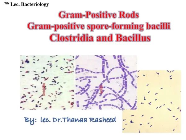
Gram-Positive BacilliPart three MLAB 2434: Microbiology Keri Brophy-Martinez
Corynebacteria • Significant Corynebacterium species • C. diphtheriae • C. xerosis • C. pseudodiphtheriticum • C. pseudotuberculosis • C. jekeium • C. ulcerans
Corynebacterium Species:General Characteristics • Found as free-living saprophytes in fresh and salt water, in soil and in the air • Members of the usual flora of humans and animals(often dismissed as contaminants) • Often called “diphtheroids” or “corneforms” • Corynebacterium diphtheriae isthe most significant pathogen • Other species may cause infections in immunocompromised hosts
Corynebacterium Species:General Characteristics • Morphology • Gram-positive, non–spore-forming rods • Arrange in palisades:“L-V” shape; “Chinese characters” • Pleomorphic: “club-ends” or coryneform • Beaded, irregular staining
C. diphtheriae: Agent of Diphtheria • Toxigenic Corynebacterium diphtheriae • Worldwide distribution but rare in places where vaccination programs exist • Exotoxin, Diphtheria toxin, as the virulence factor • Not all C. diphtheriae strains produce toxin • Disrupts protein synthesis • Triggers cell lysis
Toxigenic Corynebacterium diphtheriae • Toxin consists of two fragments • A: Active fragment • Inhibits protein synthesis • Leads to cell/tissue death • B: Binding • Binds to specific cell membrane receptors • Mediates entry of fragment Ainto cytoplasm of host cell
Clinical Forms of Diphtheria • Respiratory • Acquired by droplet spray or hand to mouth contact • Non-immunized individuals are susceptible • Non-respiratory • Systemic form • Toxin is absorbed in the blood stream and carried systemically • Affects the kidneys, heart, and nervous system • Death occurs due to cardiac failure • Cutaneous form • Seen in tropical geographic areas • Infections occur at the site of abrasions • Associated with animal contact & unpasteurized dairy products
C. diphtheriae: Causative Agent of Diphtheria • Respiratory disease–diphtheria • Incubation period–2 to 5 days • Symptoms: sore throat, fever, malaise • Toxin is produced locally, usually in the pharynx or tonsils • Toxin causes tissue necrosis, can be absorbed to produce systemic effects • Forms a tough grey to white pseudomembrane which may cause suffocation
C. diphtheriae: Causative Agent of Diphtheria • C. diphtheriae pseudomembrane • WBC + organism
C. diphtheriae:Treatment • Infected patients treated with anti-toxin and antibiotics • Anti-toxin produced in horses • Binds the circulating toxin • Antibiotics have no effect on circulating toxin, but prevent spread of the toxin • Penicillin drug of choice • DPT Immunization
Laboratory Diagnosis:Cultural Characteristics • Loeffler's slant or Pai's slant—Used to demonstrate pleomorphism and metachromatic granules ("Babes’ Ernst bodies“) • Growth on Serum Tellurite or modified Tinsdale exhibits brown or grayish→ to black halos around the colonies
Laboratory Diagnosis • Microscopic morphology • Gram-positive, non–spore-forming rods, club-shaped • Appear in palisades and give "Chinese letter" arrangement • Can be beaded • From the production of metachromatic granules Corynebacterium diphtheriae gram stain
Laboratory Diagnosis:Corynebacterium diphtheria • Identification • Confirm identification by fermentation reactions(glucose +) • Catalase positive • Urease negative • Non-motile
Laboratory Diagnosis • Toxigenicity testing • Elek test • Immunodiffusion test • Organisms are streaked on media with lox Fe content to maximize toxin production. • Identification of C.diphtheriae does NOT mean the patient has dipheria. Must show the isolate produces the toxin.
Corynebacteriumjekeium • Clinical Infections • Septicemia • Meningitis • Bacteremia • Pulmonary disease • Populations Affected • Immunosuppressed • IV drug users • Recent invasive procedure
C. jeikeiumIsolation & Identification • BAP: 48-72 hours @ 35oC in ambient air or 5% CO2 small, gray-white colony, nonhemolytic • Gram stain: pleomorphic, club-shaped GPR arranged in V forms or palisades • Key Biochemicals • Catalase= positive • Nitrate reduction= negative • Urea= negative • Sucrose= negative • Glucose= positive • Resistant to most antibiotics • Susceptible to vancomycin
Listeria monocytogenes:General Characteristics • Gram-positive, non–spore-forming rods • Only human pathogen in genus • Widespread in nature • Known to infect a wide variety of animals • Human exposure is limited; direct or indirect • Transient colonization occurs without disease
Listeria monocytogenes:Clinical Infections • Adults • Septicemia/meningitis in the compromised/elderly • Mild flu-like syndrome in pregnant womencould be fatal to fetus • Ingestion of contaminated food (cottage cheese, coleslaw, chicken, hot dogs, lunch meat) • Neonatal • Early onset from intrauterine transmission results in sepsis; high mortality rate • Late onset manifests as meningitis; lower mortality rate
Listeriamonocytogenes:Virulence Factors • Hemolysin ( Listeriolysin O) • damages host cell membrane • Superoxide dismutase • Resists toxic effects of the host • P60 surface protein • Induces phagocytosis thru adhesion and penetration
Laboratory Diagnosis: L. monocytogenes • Identification • Microscopic morphology • Gram Positive non–spore-forming coccobacillary, pairs or short chains • Colony Morphology • Grows well on blood agar; colonies produce a narrow zone of hemolysis similar to Group B Streptococcus • Small, round and translucent
Laboratory Diagnosis: L. monocytogenes • Grows well at 0.5° C to 45° C • Because of this temperature range, especially the cooler end of the range, this organism grows well in refrigerated products, such as cream, cheese, deli meats, etc. • Can sometimes be isolated after “cold enrichment” (hold broth at 4° C for several weeks and subculture)
Laboratory Diagnosis: L. monocytogenes • Identification • Catalase positive • Motility: • Motile at 25o C; "umbrella" type → • Tumbling motility in hanging drop preparations (this can be seen on Gram Stain Tutor at www.medtraining.org) “Umbrella” motility pattern (Left) typical for L. monocytogenes
Laboratory Diagnosis: L. monocytogenes • Identification • CAMP test • Produces a “block” type of hemolysis in contrast to “arrow”-shape produced by Group B Streptococcus CAMP test with Listeria monocytogenes Positive CAMP test for Group B Streptococcus
Differentiating Characteristics betweenL. monocytogenesand Other Gram Positive Bacteria
References • Engelkirk, P. G., & Duben-Engelkirk, J. (2008). Laboratory Diagnosis of Infectious Diseases: Essentials of Diagnostic Microbiology . Baltimore, MD: Lippincott Williams & Willkins. • http://en.wikipedia.org/wiki/Lactobacillus • http://www.thefullwiki.org/Corynebacterium_diphtheriae • http://quizlet.com/10262287/print/ • Kiser, K. M., Payne, W. C., & Taff, T. (2011). Clinical Laboratory Microbiology: A Practical Approach . Upper Saddle River, NJ: Pearson Education, Inc. • Mahon, C. R., Lehman, D. C., & Manuselis, G. (2011). Textbook of Diagnostic Microbiology (4th ed.). Maryland Heights, MO: Saunders.